
|
Fig. 1
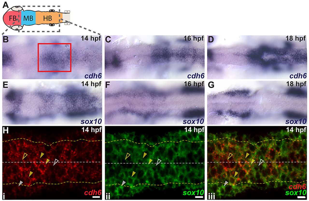
cdh6 is expressed dynamically in the hindbrain and NCCs. (A) Embryonic brain divisions. Gray box shows region displayed in B-G. (B-G) Dorsal views (anterior left) of cdh6 and sox10 in situ hybridizations at 14hpf (B,E), 16hpf (C,F) and 18hpf (D,G). Red box marks approximate area shown in H. (H) Confocal images (dorsal views, anterior left, individual z-planes) of fluorescent in situ hybridizations for cdh6 (i, iii) and sox10 (ii, iii) at 14hpf. Yellow dashed lines mark basal neuroepithelial surfaces and white dashed lines mark apical midlines. Cells between the yellow dashed lines are neuroepithelial cells or premigratory NCCs. Cells outside the yellow dashed lines are mesenchymal cells or delaminated NCCs. Closed yellow arrowheads mark premigratory NCCs expressing cdh6 and sox10. Open yellow arrowheads mark neuroepithelial cells expressing only cdh6. Open white arrowheads mark NCCs expressing only sox10. Closed white arrowheads mark NCCs outside the neuroepithelium expressing only sox10. FB, forebrain; MB, midbrain; HB, hindbrain. Scale bars: 10μm.