
|
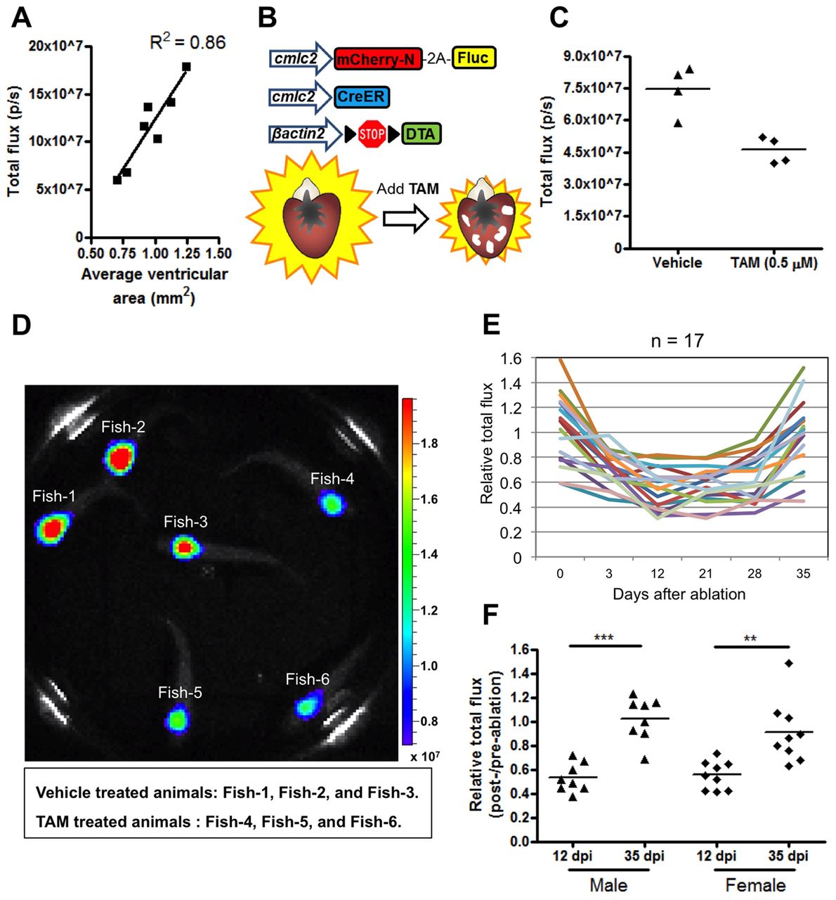
Fig. 4
Longitudinal monitoring of heart regeneration by luciferase imaging. (A) Correlation between bioluminescence (p/sec) and the surface area of tissue sections from adult ventricles. One representative experiment is shown here from two experiments. (B) Cardiac ablation/detection constructs and injury. TAM, tamoxifen. (C) Bioluminescence recorded 10 days after induced genetic cardiomyocyte ablation. One representative experiment is shown here from three replicate experiments. (D) Snapshot of a movie of six swimming adult triple-transgenic animals from these experiments, at 10 days post-treatment. Three have received tamoxifen and have lower bioluminescence signals (Fish-4, Fish-5 and Fish-6) than vehicle-treated animals (Fish-1, Fish-2 and Fish-3). Luminescence signals are reported as radiance (p/sec/cm2/sr) with a color bar. (E) Relative total flux indicating changes in bioluminescence in 17 animals during the course of injury and regeneration. (F) Relative total flux showing increases in bioluminescence between 12 days and 35 days after tamoxifen treatment. Animals are grouped here by sex. ***P<0.001, **P<0.01, paired Student’s t-test. dpi, days post-injury.