
Fig. 5
|
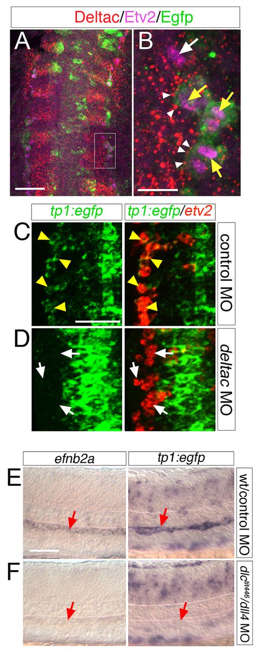
Fig. 5
deltac and dll4 have overlapping roles during artery differentiation. (A) Flat-mounted Tg(tp1:egfp)um14 embryo at 12 ss immunostained with antibodies against Deltac (red), Etv2 (magenta) and Egfp (green). Dorsal view, anterior is up. Box indicates magnified region in B. (B) White arrow denotes nucleus of Egfp-negative, Etv2-positive endothelial progenitor. Yellow arrows denote Etv2-positive nuclei of Egfp-positive cells. White arrowheads mark Deltac puncta adjacent to Egfp-positive endothelial cells. (C,D) Flat-mounted Tg(tp1:egfp)um14 embryos at 10 ss following two-color fluorescence in situ hybridization with riboprobes against egfp (green) and etv2 (red) imaged by confocal microscopy. Dorsal view, anterior is up. Yellow arrowheads denote etv2/egfp-positive cells; white arrows are etv2/egfp-negative cells. Embryos injected with 10 ng of control MO (C), or 10 ng of deltac MO (D). (E,F) DIC images of Tg(tp1:egfp)um14 embryos subjected to whole-mount in situ hybridization with riboprobes against efnb2a (left panels) or egfp (right panels). (E) Wild-type embryo injected with 15 ng control MO. (F) dlctit446 mutant embryo injected with 15 ng dll4 MO. Scale bars: 40 μm (A,C); 10 μm (B); 50 μm (E,F).