
|
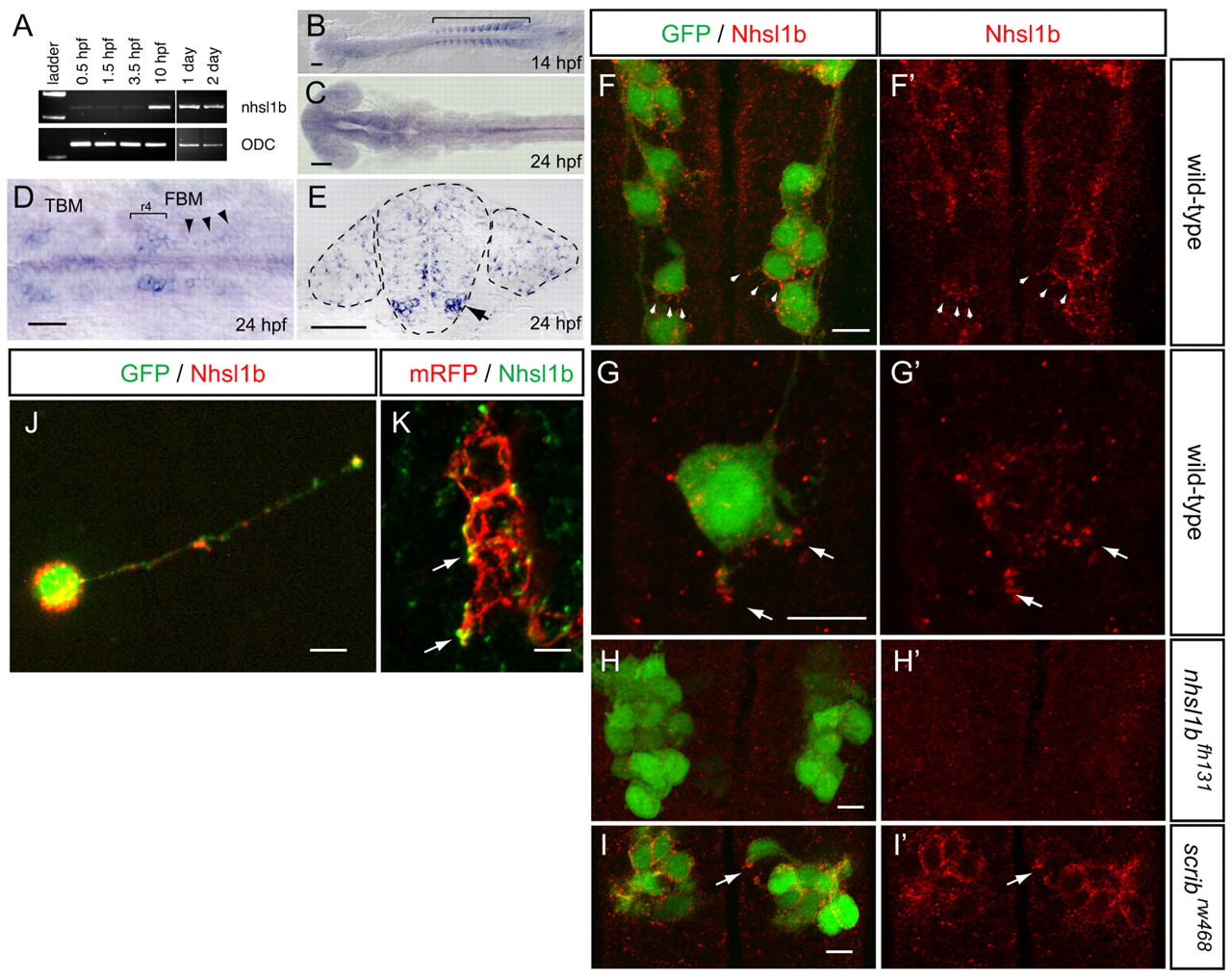
Fig. 4 nhsl1b is expressed in facial branchiomotor (FBM) neurons and localizes to membrane protrusions. (A) RT-PCR from fertilization to 2 days old shows onset of zygotic nhsl1b expression at the end of epiboly [10 hours post-fertilization (hpf)]. ODC, ornithine decarboxylase control. (B-E) mRNA in situ hybridization with nhsl1b in whole mount (B-D) and in cross section at the level of r5 (E) showing widespread, low-level expression in somites (B) and CNS (C) and specific upregulation in cranial motorneurons (D,E). nhsl1b is expressed in FBM neurons in rhombomere (r)4 at the onset of migration (bracket in D) and in r5 and r6 during migration (arrowheads in D, arrow in E) and in trigeminal branchiomotor neurons in r2 (TBM). (F-I2) Whole-mount immunocytochemistry with anti-Nhsl1b (red). Isl1:GFP marks FBM neurons (green). Nhsl1b is localized to the membrane surface of FBM neurons, particularly to membrane protrusions (arrowheads) in wild type (F,G) but not Nhsl1b mutant embryos (H). Nhsl1b is similarly localized in FBM neurons in Scrib mutants (I). I2-J2 show Nhsl1b immunostaining alone. (J) Primary cultures of FBM neurons isolated from Tg(isl1:GFP) fish immunostained for Nhsl1b (red) and GFP (green) shows colocalization of Nhsl1b in motorneurons. (K) Anti-Nhsl1b staining in Tg(isl1CREST-hsp70l:mRFP)fh1 fish showing clear colocalization of Nhsl1b with the cell membrane (arrows). Scale bars: 50 µm for B-E; 7 µm for F-K.