
Fig. 8
|
Fig. 8
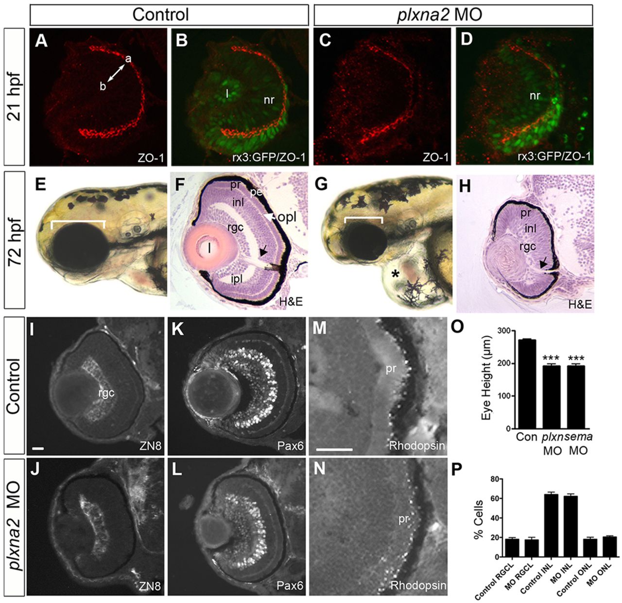
plxna2 morphants form retinas with the appropriate cell types. (A-D) Transverse sections of 21hpf control (A,B) and plxna2 (C,D) morphant rx3:GFP optic cups immunolabeled with anti-ZO-1. (B,D) Merge. a, apical; b, basal. (E-P) Analysis of 72hpf retinas. Transverse sections (F,H-N). (E-H) Control (E) and plxna2 morphant (G) indicating smaller eye (bracket) and pericardial edema (*) in the morphant that likely reflects cardiac Plxna2 expression (Toyofuku et al., 2004). (F,H) Hematoxylin and Eosin staining. Optic nerve head is indicated by arrows. (I-N) Immunolabeling of ZN8+ retinal ganglion cells (RGCs) (I,J), Pax6+ RGCs and amacrine cells (K,L), and rhodopsin+ rods (M,N). Scale bar: 50μm for I-L; 20μm for M,N. (O,P) Eye height (O) (***P<0.001; one-way ANOVA, Dunnett′s test, n=2) and % of total nuclei in each layer of the central one-third of a central retinal section (P) (control, n=6; plxna2 morphant, n=8). Error bars indicate s.e.m. inl, inner nuclear layer; l, lens; ipl, inner plexiform layer; nr, neural retina; opl, outer plexiform layer; pe, pigment epithelium; pr, photoreceptors.