
|
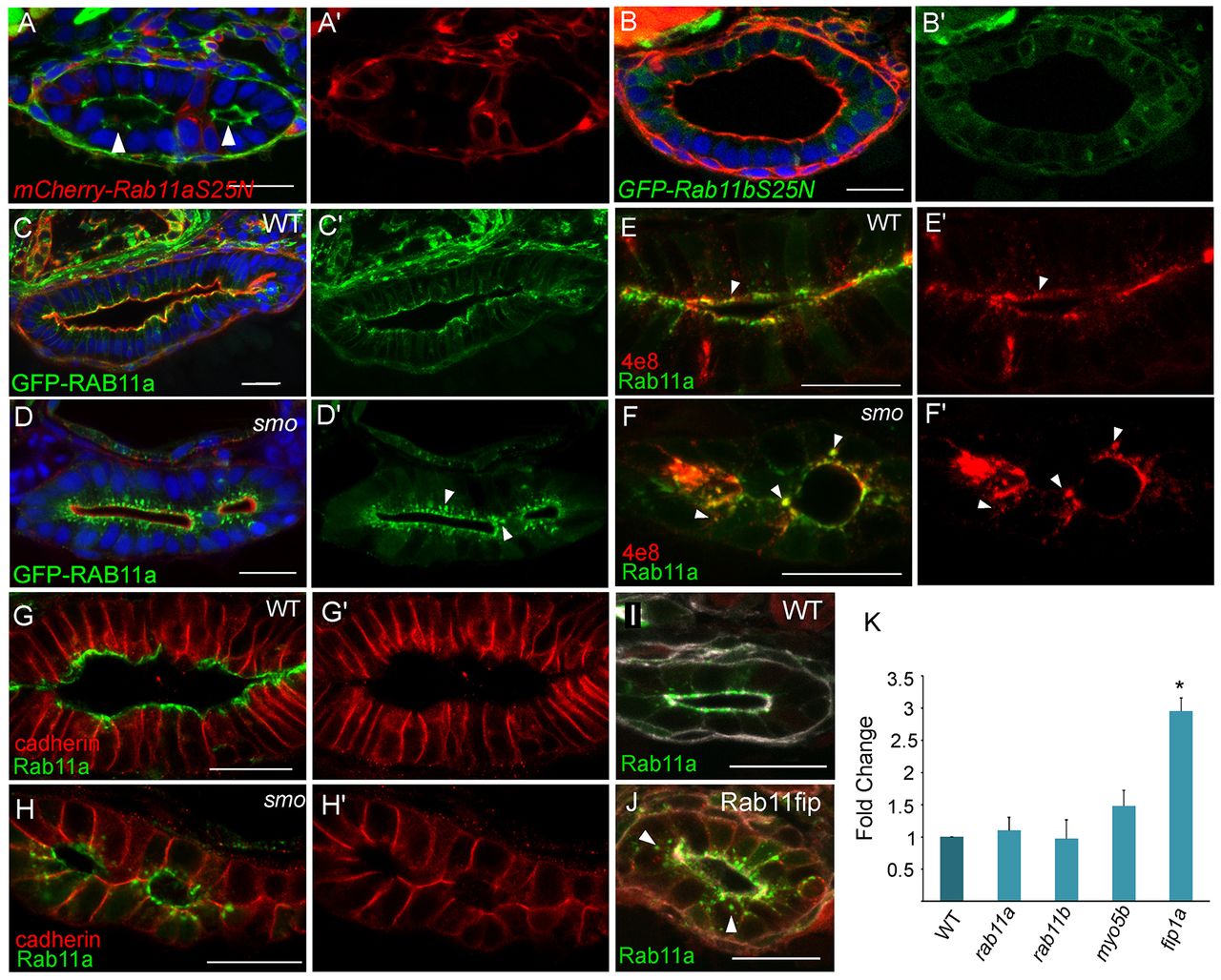
Fig. 6
Rab11 is abnormally localized in smos294 mutants. (A,A2) Confocal cross section of a Tg(hsp70l:gal4); Tg(UAS:rab11aS25N) embryo. Phalloidin (green). (B,B2) Confocal cross section of a Tg(hsp70l:rab11bS25N) embryo. Phalloidin (red) (C,D) Confocal cross sections of smos294 and wild-type clutchmates expressing Tg(hsp70l:GFP-RAB11a). Arrowheads point to abnormal enlarged Rab11 positive vesicles in smos294. Phalloidin (red) (E-H) Confocal cross section of wild-type and smos294 embryos expressing Tg(hsp70l:GFP-RAB11a) stained for the apical marker 4e8 (E,F) or cadherin (G,H). Arrowheads point to areas of colocalization. (I,J) Confocal cross sections of uninjected and RFP-Rab11fipa1-injected Tg(hsp70l:GFP-RAB11a) embryos. Arrowheads point to dispersed compartments. (K) Expression levels of Rab11 family members in the intestinal epithelium of smos294 mutants relative to wild-type clutchmates. Rab11fip1a P<0.011, n=3. All embryos are 72 hpf. Scale bars: 20 μm.