
|
Fig. 2
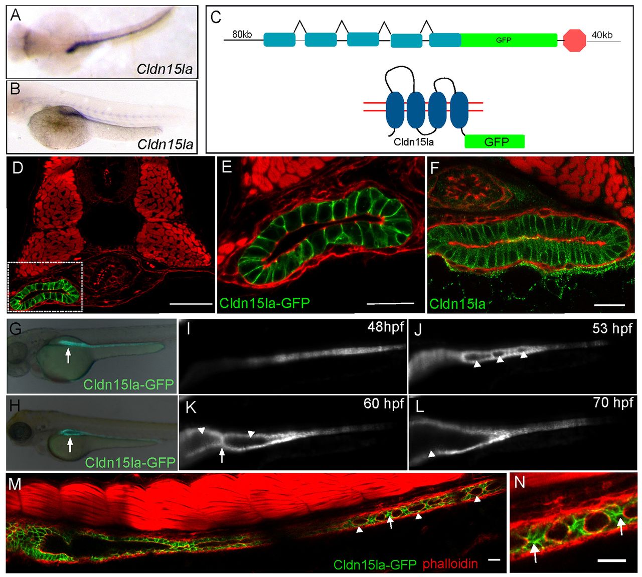
Generation of an intestine-specific transgenic line. (A,B) Dorsal, top panel, and lateral view, bottom panel, in situ hybridization showing claudin 15-like a expressed specifically in the intestine at 56 hpf. (C) Schematic of TgBAC(cldn15la-GFP) generation. The recombination target is shown on top, and the expected protein structure is shown on the bottom. (D) Confocal cross-section of a 72 hpf TgBAC(cldn15la-GFP) embryo. (E) Magnification of box from D. (F) Immunolocalization of Cldn15la to the basolateral membranes of intestinal epithelial cells. (G,H) Whole-mount fluorescent images of 55 hpf and 75 hpf embryos expressing TgBAC(cldn15la-GFP). Arrows indicate the gut tube. (I-L) Live imaging of TgBAC(cldn15la-GFP) using SPIM. Snapshots from a single plane from 48-70 hpf. Arrowheads indicate lumens; arrows point to cell-cell contacts between lumens. (M) Stitched confocal whole-mount images of a TgBAC(cldn15la-GFP) embryo show unfused lumens (arrowhead) in the posterior intestine at 68 hpf that are separated by cell-cell contacts (arrow). (N) Magnification of cell-cell contacts from M. Arrows indicate contacts. Phalloidin (red). Scale bars: 50 μm in D-F; 20 μm in M,N.