
Fig. S5
- ID
- ZDB-IMAGE-140716-5
- Publication
- Nguyen-Chi et al., 2014 - Transient infection of the zebrafish notochord with E. coli induces chronic inflammation
- All Figures
- Figures for Nguyen-Chi et al., 2014
|
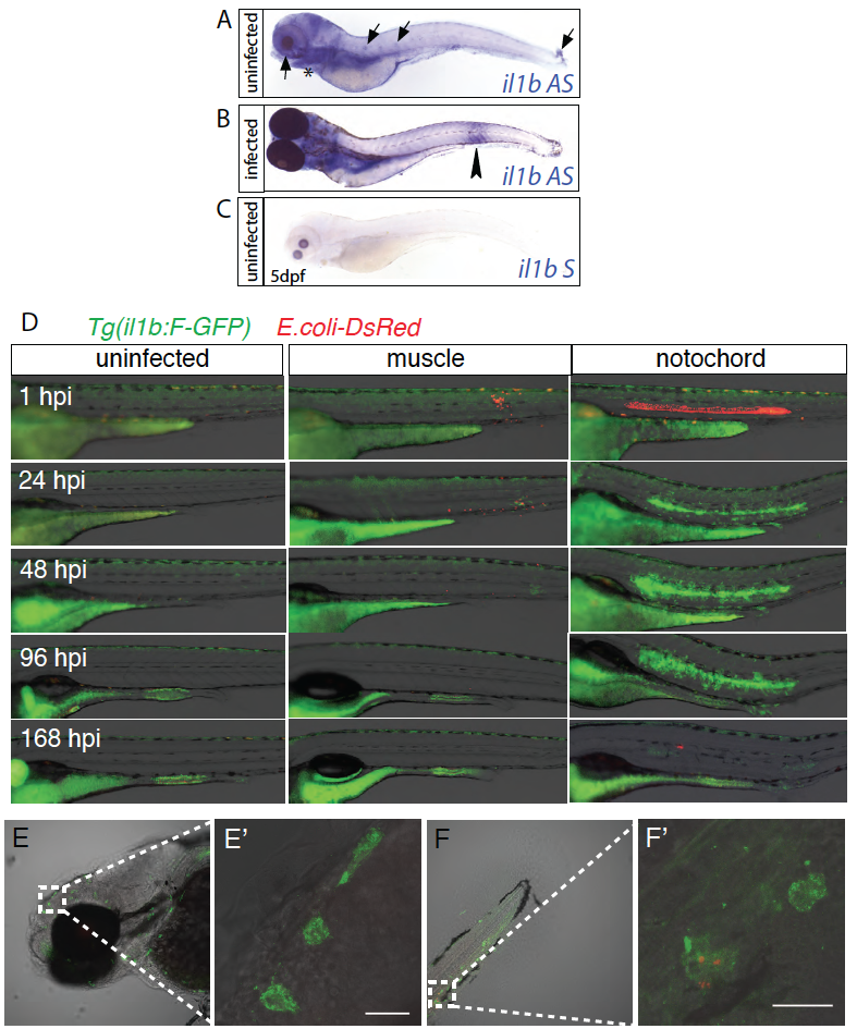
Fig. S5
transcriptional activation of il1b upon infection with E. coli
(A-B) Whole mount in situ hybridization with il1b anti-sense (il1b AS) probe in 5dpf larvae uninfected (A) and infected with E. coli in the notochord (3dpi) (B). Probe signal is detected in the tip of the caudal fin, the skin, the neuromast, eyes (arrows) and gills (asterisks). After infection, larvae overexpress il1b mRNA in the inflamed region (arrowhead). (C) No signal is detected using il1b sense probe (il1b S) (C). (D) GFP (green) expression in Tg(il1b:GFP-F) larvae. Larvae were imaged without infection or at different time points following infection with a DsRed expressing E. coli (red) in the muscle or in notochord. Each column shows images from a single larva image repeatedly. (E-F) Confocal analysis of GFP expression in Tg(il1b:-GFP-F) in the head (G) and the tail (H) regions 24h following E. coli injection. Maximum projections, lateral views, scale bars=20μm. (E′) and (F′) are high magnification images of regions boxed in E and F, respectively.