
Fig. S3
- ID
- ZDB-IMAGE-140716-3
- Publication
- Nguyen-Chi et al., 2014 - Transient infection of the zebrafish notochord with E. coli induces chronic inflammation
- All Figures
- Figures for Nguyen-Chi et al., 2014
|
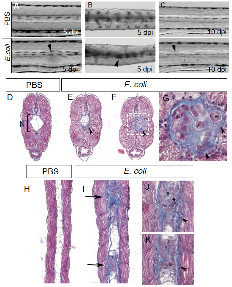
Fig. S3
Notochord infection induces notochord defects
Bright-field images of PBS or E. coli injected larvae in the notochord at 5 dpi (A-B) and 10 dpi (C). A, C lateral views; B, dorsal view. Arrowheads show defects in the notochord anterior of the injection site (5 to 10 somites away). (D-G) 4 μm cross sections of PBS (D) or E. coli (E-G) injected larvae stained with Masson’s Trichrome. While PBS injected larvae present a notochord (N, bracket) filled mainly with 2 or 3 big vacuolated cells (white) and delimited with a regular, unaffected collagen membrane (blue), E. coli injected larvae present several abnormalities of the notochord. Numerous cells and vacuoles are found inside the notochord. Increase of collagen deposition appears inside and outside the notochord (arrowhead). Many cells are embedded inside the collagen sheath (asterisks). (G) is a high magnification image of the region boxed in (F). (H-K) 4 μm longitudinal sections of PBS (H) or E. coli (I-K) infected larvae stained with Masson’s Trichrome. As described above, PBS injected larvae present a notochord filled mainly with vacuolated cells (H), while E. coli infected larvae present several massive cell aggregates within the notochord (I). In these locations, the notochord is surrounded with cells embedded in the matrix (arrow heads) and cells with different morphologies are found inside the notochord (* and #) (J-K).