
|
Fig. 4
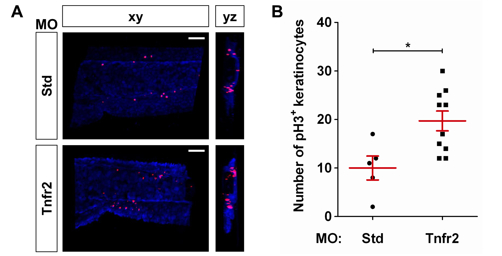
Tnfr2 deficiency results in increased proliferation of skin keratinocytes.
Zebrafish one-cell krt18:RFP embryos were injected with standard control (Std) or Tnfr2 MOs. (A) Representative frontal (xy) and lateral (yz) tridimensional reconstructions from confocal microscopy images of WIHC of krt18:RFP larvae stained at 3 dpf with anti-RFP (keratinocytes, blue) and anti-phosphorylated H3 (pH 3, proliferation marker) antibodies. (B) Quantification of the number of pH3/RFP+ (i.e., proliferating keratinocytes) cells in the CHT area. Each dot represents one single larva, and the mean ± S.E.M. for each group of larvae is also shown. Scale bars, 100 μm. *p<0.05.