
|
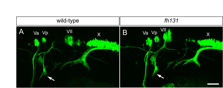
Fig. S1 Cranial nerve architecture in nhsl1b mutants. Live confocal images of isl1:GFP transgene expression in lateral view in wild-type (A) and nhsl1bfh131 mutant (B) embryos at 48 hours post-fertilization (hpf). Va and Vp, anterior and posterior nuclei, respectively, of the fifth cranial nerve (trigeminal); VII, nucleus of the seventh cranial nerve (facial), which is migrated into r6 in wild-type and is unmigrated in mutant embryos; X, nucleus of the tenth cranial nerve (vagus). Arrows indicate the projection of the facial nerve into the second pharyngeal arch, which is unaffected in nhsl1bfh131 mutants in spite of the failure of the FBM neurons to migrate out of r4. Scale bar: 50 µm.