
Image
|
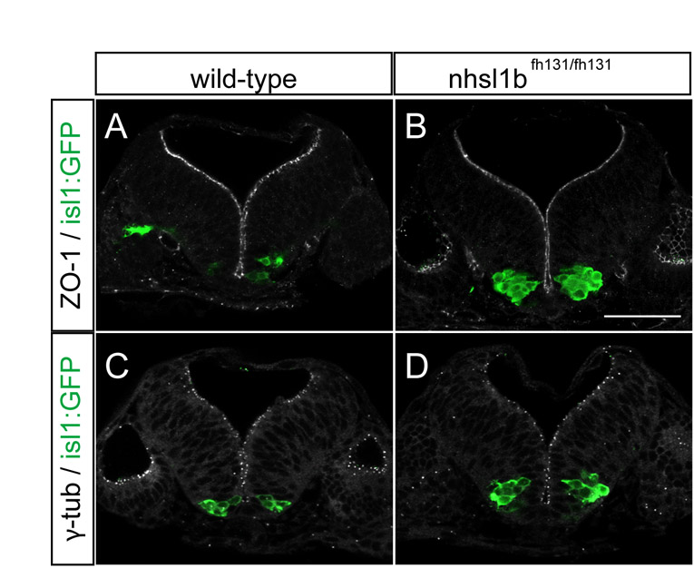
Figure Caption
Fig. S5 Nhsl1b is not required for neuroepithelial apico-basal polarity. (A-D) Confocal images of cross sections at the level of rhombomere (r)4 showing normal apico-basal polarity in the neuroepithelium of 24 hours post-fertilization (hpf) wild-type (A,C) and nhsl1bfh131 mutant (B,D) embryos. ZO-1 marks sub-apical tight junctions of progenitors (A,B) and γ-tubulin marks apical centrosomes (C,D). isl1:GFP marks FBM neurons which accumulate in r4 in nhsl1bfh131 mutants (B,D). Scale bar: 50 µm.
Acknowledgments
This image is the copyrighted work of the attributed author or publisher, and
ZFIN has permission only to display this image to its users.
Additional permissions should be obtained from the applicable author or publisher of the image.
Full text @ Development