
|
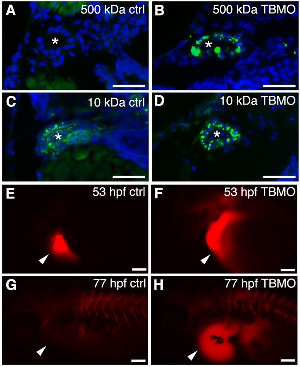
Fig. 4
Knockdown of sept7b affects glomerular barrier function and fluid flow. (A,B) 500-kDa fluorescein dextran (green) injected into the cardinal vein of 3.5-dpf larvae passes the glomerular barrier and is endocytosed into the pronephric tubules of sept7b-TBMO-injected larvae (B), whereas in the control (ctrl; A) the dextran remains in circulation. (C,D) 10-kDa fluorescein dextran readily passes the glomerular barrier in both control (C) and TBMO-injected (D) larvae and is endocytosed into the pronephric tubules. DAPI (blue) stains the nuclei, and the asterisk marks the lumen of the tubule. (E–H) Tetramethylrhodamine dextran (70,000-kDa) was injected into the pericardial sac (arrowheads) of control (E) or sept7b-TBMO-injected (F) embryos at 53 hpf that were imaged immediately after dye injection (E,F) and again at 77 hpf (G,H). Clearance of the dye was observed in the control larvae (G) but not in sept7b morphants (H). Scale bars: 20μm (A–D); 200μm (E–H).