
|
Fig. 1
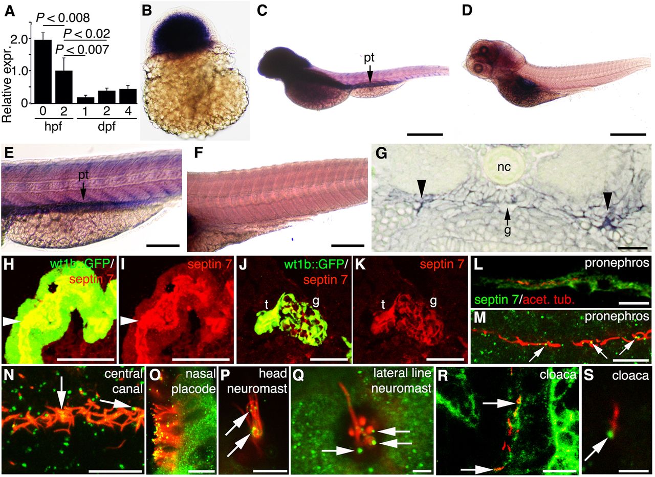
Septin 7 is expressed throughout zebrafish development and is enriched in the brain and pronephros. sept7b mRNA expression in zebrafish larvae (A–E). (A) By using qRT-PCR and normalizing to rps3 (ribosomal protein subunit 3), it can be seen that sept7b is expressed in fertilized embryos at 0 hpf, 2 hpf and in 1–4-dpf embryos and larvae. (B) In situ hybridization shows that sept7b is expressed in 3-hpf embryos. (C–F) Lateral views of 4-dpf larvae hybridized with sept7b antisense (C,E) and sense (D,F) probes. (C,E) sept7b mRNA is concentrated in the brain and the pronephric tubule (pt). (D,F) The sense probe shows no signal, except in the swim bladder region (D). (G) A section of a 2-dpf embryo shows sept7b mRNA in the glomerulus (arrow labeled g) and the pronephric tubule (arrowheads). Notochord, nc. (H–K) The septin 7 protein is localized in the pronephric tubule and glomerulus. (H,I) Immunostaining shows that septin 7 (red) localizes in the pronephric tubule cells (t; arrowhead), and in the glomerulus (g) of 4-dpf wt1b::GFP zebrafish larvae (J,K). (L–S) Septin 7 is expressed in ciliated cells in 72-hpf embryos. Cilia are visualized by staining for acetylated tubulin (red). Septin is indicated by green fluorescence. (L) Septin 7 localizes in pronephric tubule cells. (M) In addition to localizing in the cytoplasm of pronephric tubule cells, septin 7 concentrates at the base of or along the cilia (arrows). (N) Septin 7 is found at the base of, or along, the cilia in the central canal (arrows). (O) Septin 7 is expressed in nasal epithelial cells, along the ciliary axoneme and in the tips of cilia. (P) Septin 7 is localized along the axoneme of the neuromast in the head region (arrows). (Q) In lateral line neuromasts, septin 7 is found close to the base of cilia (arrows). (R) In the cloaca region, septin 7 is found in the epithelium and at the base of cilia (arrows). (S) Higher magnification reveals a concentration of septin 7 at the base of cilia in cloaca (arrow). Scale bars: 100μm (B,E,F); 300μm (C,D); 20μm (G,R); 50μm (H,I); 40μm (J,K); 10μm (L–O); 7.5μm (P); 2.5μm (Q); 3μm (S).