
|
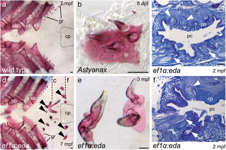
Fig. 4
The upper of pharynx of adult zebrafish is only partially restored to the ancestral condition by ectopic eda expression. (A and D) Dorsal views of alizarin-stained pharyngeal arches with fifth ceratobranchials removed to show location of ectopic teeth (arrowheads in D) posterior to upper gill arch elements and dorsal to fifth ceratobranchials. Approximate position of the chewing pad is indicated by dotted lines. (E) Calcified tissue uniting ectopic teeth resembles upper pharyngeal toothplates of A. mexicanus (B). (C and F) Transverse sections (approximate plane of section indicated in D) reveal teeth in the vicinity of the palatal organ and projecting into the pharyngeal cavity (arrowheads in C) as well as lateral to the chewing pad (arrowhead in F). Lower pharyngeal teeth in (C and F) indicated by arrows. Abbreviations: cb, ceratobranchial; cp, chewing pad; eb, epibranchial; gr, gill raker; mpf, months postfertilization; pb, pharyngobranchial; pc, pharyngeal cavity; po, palatal organ; tp, toothplate. (Scale bars, 50 μm.)