
Image
|
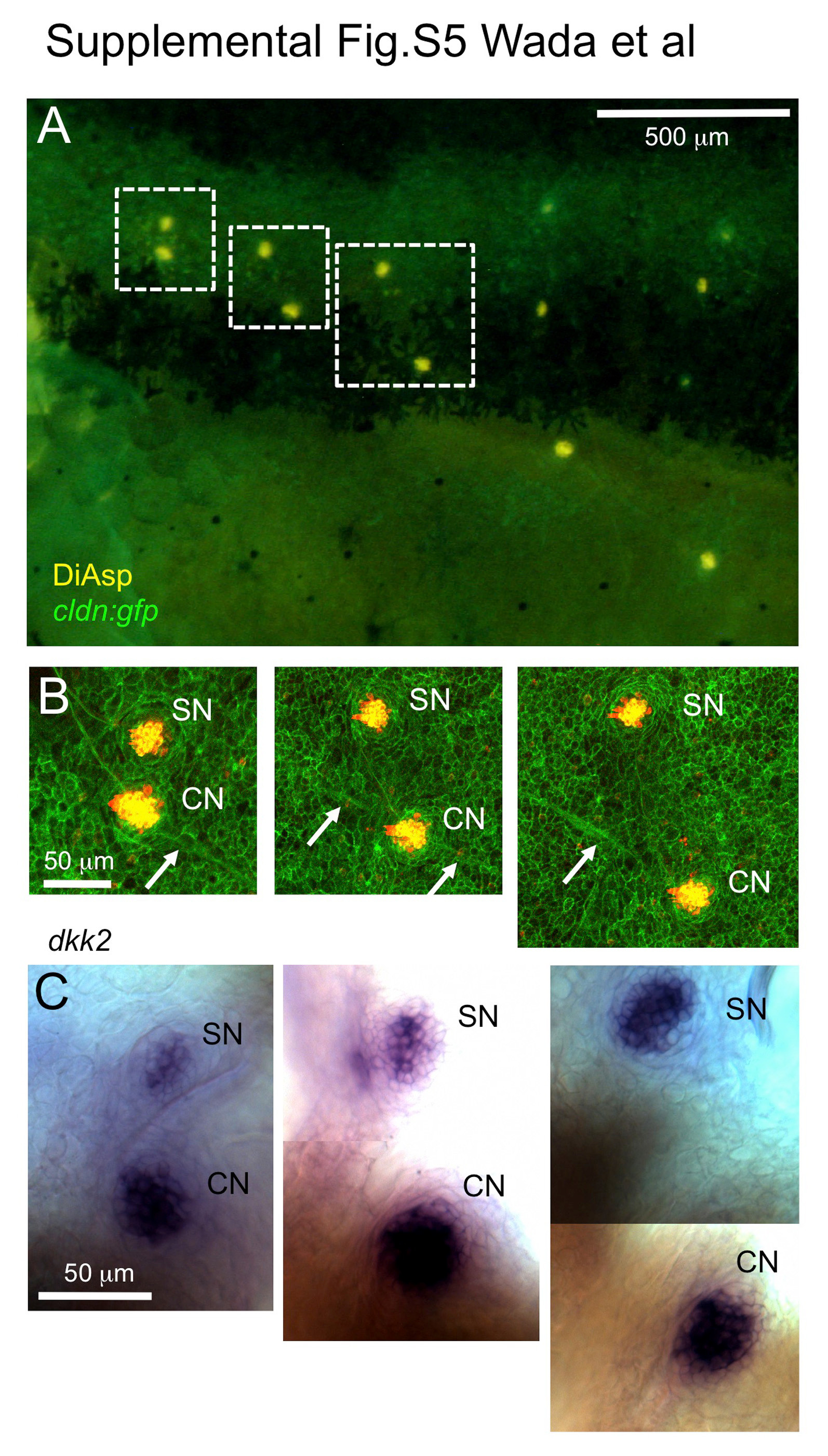
Figure Caption
Fig. S5
dkk2 is expressed in superficial neuromasts and presumptive canal neuromasts. (A) A 10 mm SL cldn:gfp larva stained with DiAsp. (B) Higher magnification of the boxed region in A. Arrows indicate interneuromast cells. SN, superficial neuromast; CN, canal neuromast. (C) The same larva was stained with a dkk2 RNA probe (Wada et al., 2013).
Acknowledgments
This image is the copyrighted work of the attributed author or publisher, and
ZFIN has permission only to display this image to its users.
Additional permissions should be obtained from the applicable author or publisher of the image.
Reprinted from Developmental Biology, 392(1), Wada, H., Iwasaki, M., Kawakami, K., Development of the lateral line canal system through a bone remodeling process in zebrafish, 1-14, Copyright (2014) with permission from Elsevier. Full text @ Dev. Biol.