
Fig. S4
- ID
- ZDB-IMAGE-140701-21
- Publication
- Wada et al., 2014 - Development of the lateral line canal system through a bone remodeling process in zebrafish
- All Figures
- Figures for Wada et al., 2014
|
Fig. S4
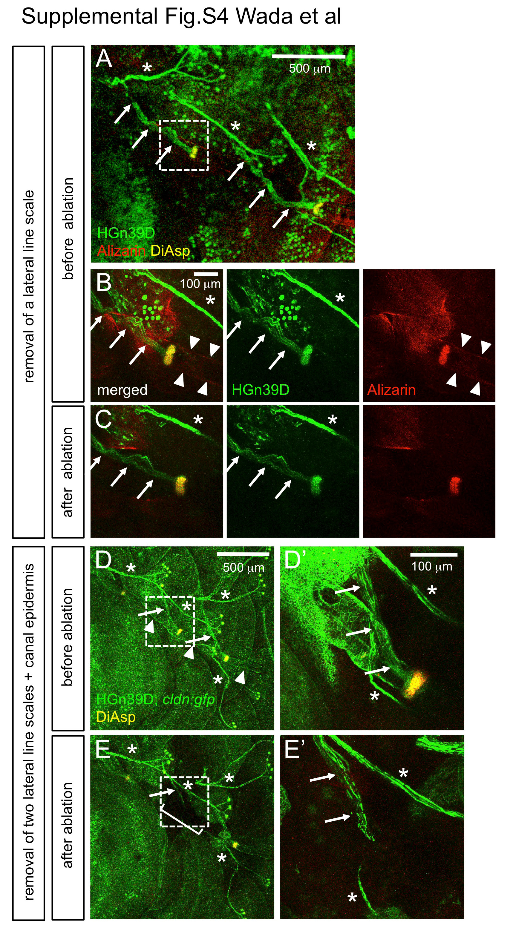
Axonal projections were not affected by scale removal. (A) The lateral line nerves are visualized in HGn39D enhancer trap line (Faucherre et al., 2009) stained with DiAsp and Alizarin red. Branches of lateral line nerves innervating canal neuromasts are indicated by arrows. (B) Higher magnification of the boxed region in A. Arrowheads indicate canal roof stained with Alizarin red. (C) The lateral line scale was removed from the same fish. The canal neuromast and the lateral line nerve remain intact after scale removal. (D) The lateral line nerves and canal epidermis were visualized in HGn39D; cldn:gfp double transgenic fish stained with DiAsp. (E) Two consecutive lateral line scales and the canal epidermis are removed from the same fish (bracket). The lateral line nerve innervating the canal neuromast remains intact near the ablated site. (D′, E′) Higher magnification of the boxed region in D and E. Asterisks indicate the nerves innervating superficial neuromasts.
Reprinted from Developmental Biology, 392(1), Wada, H., Iwasaki, M., Kawakami, K., Development of the lateral line canal system through a bone remodeling process in zebrafish, 1-14, Copyright (2014) with permission from Elsevier. Full text @ Dev. Biol.