
Image
|
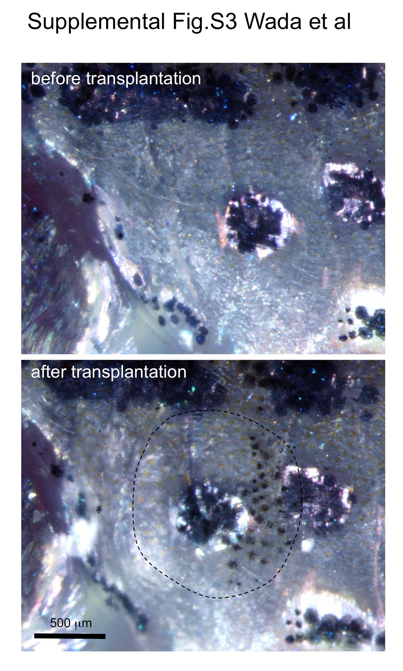
Figure Caption
Fig. S3
Scale transplantation experiments. First, one dorsal scale and one lateral line scale were removed. The dorsal scale was then inserted into the place where the lateral line scale had just been removed as described previously (Shinya and Sakai, 2011). Dorsal scales contain melanophores, whereas lateral line scales are nonpigmented. Thus we could assess whether the transplanted scale was retained by the presence of pigment cells. The transplanted scale is outlined by the dotted line. Images were taken under light microscopy using epi-illumination.
Acknowledgments
This image is the copyrighted work of the attributed author or publisher, and
ZFIN has permission only to display this image to its users.
Additional permissions should be obtained from the applicable author or publisher of the image.
Reprinted from Developmental Biology, 392(1), Wada, H., Iwasaki, M., Kawakami, K., Development of the lateral line canal system through a bone remodeling process in zebrafish, 1-14, Copyright (2014) with permission from Elsevier. Full text @ Dev. Biol.