
Image
|
Figure Caption
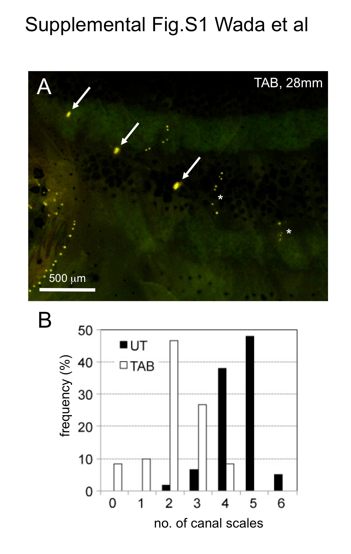
Fig. S1
Lateral line scale numbers (i.e., the number of canal neuromasts) differ significantly among zebrafish strains. (A) Distribution of trunk canal neuromasts in the TAB strain stained with DiAsp. Asterisks indicate lines of superficial neuromasts. (B) Difference in numbers of canal scales between UT and TAB strains. Lateral line scales were counted in UT (n = 60, both sides from 30 fish, 28.0-34.0 mm SL) and TAB strains (n = 60, both sides from 30 fish, 27.0-32.0 mm SL).
Acknowledgments
This image is the copyrighted work of the attributed author or publisher, and
ZFIN has permission only to display this image to its users.
Additional permissions should be obtained from the applicable author or publisher of the image.
Reprinted from Developmental Biology, 392(1), Wada, H., Iwasaki, M., Kawakami, K., Development of the lateral line canal system through a bone remodeling process in zebrafish, 1-14, Copyright (2014) with permission from Elsevier. Full text @ Dev. Biol.