
|
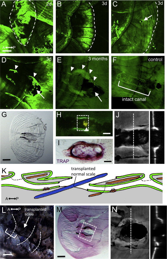
Fig. 7
The canal roof was only partly regenerated. (A–F) Fish stained with Calcein at two days (A), three days (C–D), and three months (E) after scale removal. An intact canal roof is shown in F. (G) The regenerated scale was removed and imaged by transmitted light microscopy. (H) Regenerated lateral line scale stained with DiAsp and Calcein, showing the canal neuromast attached to the removed scale. (I) TRAP staining showed abnormal osteoclast activity in the regenerated scale (intact canal is shown in Fig. 5C). (J) Confocal image of the area indicated by dotted lines in H. Optical cross sections are shown in the right panel. (K) Schematic illustration of the scale transplantation experiment. A lateral line scale was replaced by a normal scale (blue). (L) Three months after transplantation, the canal lumens were again connected (bracket) through the transplanted scale (indicated by arrow). (M) The inserted scale was removed and imaged as in G. (N) Confocal image of the area indicated by dotted lines in M. An optical cross sections is shown in the right panel. Scale bars: 100 μm in A–G, L and M, 50 μm in H and I.
Reprinted from Developmental Biology, 392(1), Wada, H., Iwasaki, M., Kawakami, K., Development of the lateral line canal system through a bone remodeling process in zebrafish, 1-14, Copyright (2014) with permission from Elsevier. Full text @ Dev. Biol.