
|
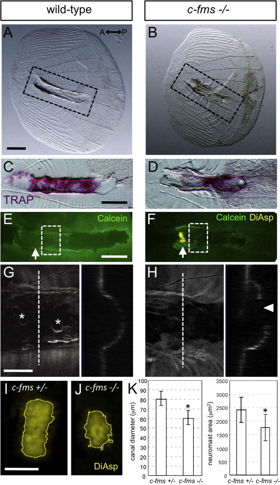
Fig. 5
Bone remodeling is required for the normal development of canal structure. (A and B) Images showing disorganization of the canal roof and (C and D) reduced TRAP activity in c-fms/panther fish. Corresponding areas are shown in dotted lines in A and B. (E and F) Removed lateral line scales stained with DiAsp and Calcein. Arrows indicate the positions of canal neuromasts, which was attached to the removed scale in c-fms/panther fish. (G and H) Confocal images of the area indicated by dotted lines in E and F. Optical cross sections are shown in the right panels. Asterisks indicate osteoclast resorption pits on the canal roof, which is not enclosed at the apical region in the c-fms/panther fish (indicated by arrowhead in H). (I and J) Growth of the canal neuromasts was perturbed in the c-fms/panther fish. Canal neuromasts stained with DiAsp were outlined using the “Find Edges” function with ImageJ software. (K) Canal diameter and neuromast area were measured in c-fms/panther fish and heterozygous siblings (28.0–32.0 mm SL). Mean±SEM is indicated. *P<0.001 (t test). Scale bars: 100 μm in A–F, 50 μm in G and I.
Reprinted from Developmental Biology, 392(1), Wada, H., Iwasaki, M., Kawakami, K., Development of the lateral line canal system through a bone remodeling process in zebrafish, 1-14, Copyright (2014) with permission from Elsevier. Full text @ Dev. Biol.