
|
Fig. 1
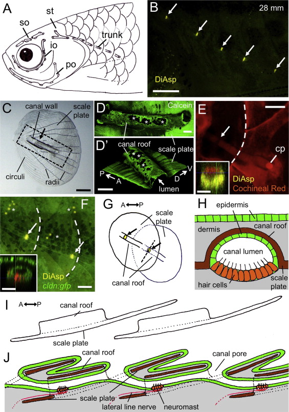
Structure of the lateral line scale in adult zebrafish. (A) Schematic distribution of canals in the UT fish strain, indicating the supraorbital (so), infraorbital (io), preopercular (po), supratemporal (st), and trunk canals. Canals were visualized experimentally by the application of black ink. In 29 out of 60 cases (48%), there were five lateral line scales, as depicted in the diagram. (B) Distribution of trunk canal neuromasts (indicated by arrows) in UT strain fish stained with DiAsp. (C) Structure of lateral line scale. Arrow indicates the position of a canal neuromast. (D and D2) Canal roof stained with Calcein. The region shown corresponds to the dotted line in C. A three-dimensional image is shown in D2. Osteoclast resorption pits are indicated by asterisks. (E and F) Canal structures are visualized with Cochineal Red staining (E), and by using the cldn:gfp line (F). Arrows, canal neuromasts; asterisks, superficial neuromasts; cp, canal pore. Caudal margins of scales are indicated by dotted lines. Optical cross sections at the level of canal neuromasts are shown in insets. (G) Schematic drawing representing a junction of two consecutive lateral line scales. Arrows indicate canal neuromasts. (H) Schematic drawing of the canal structure in cross section at the position of canal neuromasts, as indicated by the dotted line in G. (I) Lateral view showing two consecutive lateral line scales. (J) Schematic drawing of a longitudinal section showing the relationship of the canal lumen to the lateral line scales. Color used in diagrams: green, epidermis; gray, dermis; brown, ossified scale; orange, neuromast hair cells; red, lateral line nerve. Scale bars: 500 μm in B, 100 µm in C, E, and F, 20 μm in D and D2, insets in E and F.
Reprinted from Developmental Biology, 392(1), Wada, H., Iwasaki, M., Kawakami, K., Development of the lateral line canal system through a bone remodeling process in zebrafish, 1-14, Copyright (2014) with permission from Elsevier. Full text @ Dev. Biol.