
|
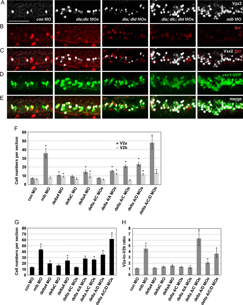
Fig. 3 Mib, DeltaA, DeltaC, and DeltaD, but not Delta4 play important roles in V2 development. (A–E) V2a and V2b cells were identified as vsx1:GFP/Vsx2 double positive and vsx1:GFP/Scl double positive cells, respectively. Side views of embryos (control MO, deltaA/deltaC double MOs, deltaA/deltaD double MOs, deltaA/deltaC/deltaD triple MOs, or mib MO) at 24 hpf with anterior to the left, dorsal up. Bar scale: 50 µm. (F) V2a and V2b cell numbers per section, (G) total V2 cell number (V2a+V2b) per section, and (H) V2a-to-V2b ratio in mib and delta knockdown embryos. (F–H) Control MO, n=21; mib MO, n=4; deltaA MO, n=23; deltaC MO, n=4; deltaA MO, n=5; delta4/deltaA MOs, n=25; delta4/deltaC MOs, n=14; deltaA/deltaC MOs, n=27; deltaA/deltaD MOs, n=20; deltaA/deltaC/deltaD MOs, n=8. Asterisks indicate statistically significant differences relative to the control (p<0.01). Error bars, SEM.
Reprinted from Developmental Biology, 391(2), Okigawa, S., Mizoguchi, T., Okano, M., Tanaka, H., Isoda, M., Jiang, Y.J., Suster, M., Higashijima, S.I., Kawakami, K., Itoh, M., Different combinations of Notch ligands and receptors regulate V2 interneuron progenitor proliferation and V2a/V2b cell fate determination, 196-206, Copyright (2014) with permission from Elsevier. Full text @ Dev. Biol.