
|
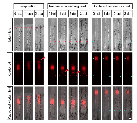
Fig. S3 Dedifferentiated osteoblasts migrate to the site of injury. Individual entpd5:Kaede fish were repeatedly imaged at the indicated time points. Left: In response to fin amputation, photoconverted osteoblasts of entpd5:Kaede transgenic fish (red) migrate to and beyond the amputation plane (arrowhead) as seen by repeated imaging of the same fish. Middle: Photoconverted osteoblasts in a segment adjacent to a fracture migrate towards the fracture (arrowhead). Right: Photoconverted osteoblasts in the second segment from the fracture do not change position. Red and blue dotted lines, fracture. Scale bar, 100 µm.