
|
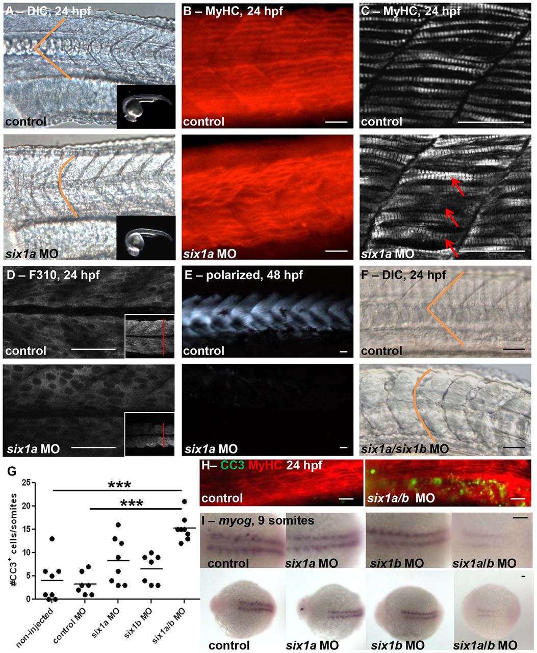
Fig. 2 six1a and six1b function together during zebrafish myogenesis. Morpholino (MO)-mediated knockdown of six1a results in disrupted U-shaped versus normal V-shaped (indicated in orange) somite morphology as observed at 24hpf by (A) DIC microscopy (control, n = 35/35; six1a MO, n = 60/83; the inset images demonstrate matched staging of embryos) and (B) Myosin Heavy Chain (MyHC) immunofluorescence (control, n = 7/10; six1a MO, n = 10/16). (C–E) There is disarrayed muscle fiber organization upon six1a knockdown as revealed by confocal imaging of (C) both fast and slow MyHC with A4.1025 antibody (arrows indicate crossover of fibers), (D) fast-muscle-specific fibers with F310 antibody (inset demonstrates decrease in cross-sectional diameter) at 24 hpf, and (E) polarized-light-mediated detection of birefringence (control, n = 17/17; six1a MO, n = 13/19) of embryos at 48 hpf. (F) Dual MO-mediated knockdown of both six1a/b leads to dramatically U-shaped somites at 24 hpf as shown by DIC microscopy (control, n = 75/77; six1a/six1b MO, n = 100/154), and (G,H) increased cleaved caspase-3 (CC3) immunofluorescence staining at 24 hpf. Non-injected, n = 35 total embryos; six1a MO, n = 30; six1b MO, n = 18; six1a/b MO, n = 22. ***P<0.0001, ANOVA with Bonferroni′s post-hoc test. The line represents the mean. (H) CC3 (green) in embryos double-stained with MyHC (red) to observe the muscle fibers. (I) Dorsal view of in situ hybridization at 9 somites shows an early delay of myogenin (myog) expression (control, n = 15/18; six1a MO, n = 16/19; six1b MO, n = 21/27; six1a/b MO, n = 8/17), which is strongest in the double six1a/b knockdown. Scale bars: 50µm. Anterior is to the left in all images. For A–F, H and I, n = the number of embryos represented by each image/the total number of embryos analyzed.