
|
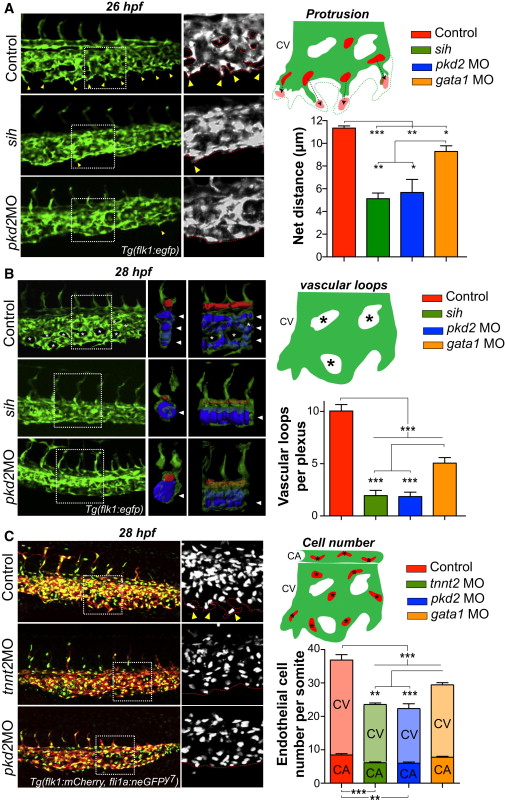
Fig. 4
Early Hemodynamics Control Embryonic Angiogenesis
(A) The protrusive activity of the CV was assessed in controls, sih mutants, and pkd2 morphants at 26 hpf. Yellow arrowheads point to vascular sprouts of the CV. Note the absence of such sprouts in both the sih mutants and pkd2 morphants. Using the Tg(fli1a:neGFP)y7 transgenic line, we tracked the nuclei of ECs at the migration front of the CV (scheme) and followed their movement over a 1 hr period. See also Movie S18. The graph shows the average net distance covered by the tracked cells in control, mutant (sih), and morphant (pkd2 and gata1) embryos (between three and four embryos were imaged per condition).
(B) Vascular morphogenesis of the CV was assessed in controls, sih mutants, and pkd2 morphants at 28 hpf. Asterisks point to vascular loops, which are mostly absent in sih mutants and pkd2 morphants (see scheme). 3D models of the vascular lumens of a delimited region (two somites) of the caudal plexus were created and are displayed as orthogonal and side views. The CA is colored in red and the CV is in blue. Images are representative of three to eight embryos imaged per condition. See also Movie S19. Arrowheads point to distinct vessels of the CV. Note that a single vessel is observed in both sih mutants and pkd2 morphants. The graph shows the number of vascular loops present in the CV at 28 hpf.
(C) The overall number of ECs in the caudal plexus (CA+CV) was assessed in controls and tnnt2a and pkd2 morphants at 28 hpf. Yellow arrowheads point to sprouting regions of the CV. Cell number was quantified in the caudal plexus of 28 hpf embryos and normalized per somite (between 6 and 11 embryos were imaged per condition; CA/V, caudal artery/vein).
Error bars depict SEM. Statistical significance was determined by unpaired Student’s t test; p < 0.05, p < 0.01, p < 0.001.