
|
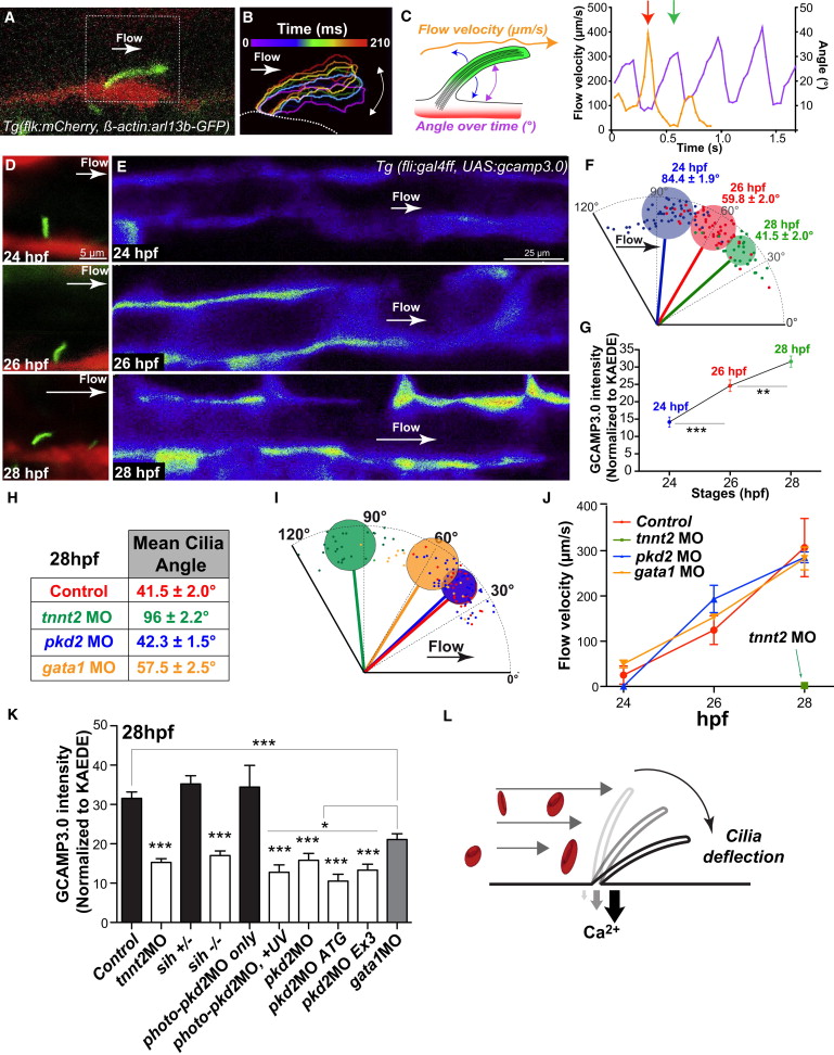
Fig. 3
Blood Flow Onset Is Mechanically Detected by Primary Cilia and Transduced via a pkd2-Dependent Calcium Increase
(A) High-speed confocal imaging at 29 fps and digital tracking in a Tg (flk1:mCherry; β-actin:Arl13b-egfp) embryo at 28 hpf. Note the cilium deflection and flow direction (white arrow). See also Movies S13 and S14.
(B) Digital tracking permits the segmentation of cilium outlines. Consecutive outlines were color-coded over time during one deflection phase (210 ms).
(C) Cilia deflection angle and flow velocity (see scheme) were quantified overtime and plotted. Note the correlation between maximum and minimum flow velocity and maximum and minimum flow deflection, respectively (red and green arrows).
(D) Cilia deflection in the DA of 24, 26, and 28 hpf embryos.
(F) Quantification of (D).
(E and G) Calcium content in the developing DA was evaluated and quantified using the endothelial-specific expression of GCAMP3.0 in Tg(fli:gal4FF; UAS:gcamp3.0) embryos. GCAMP3.0 intensity was normalized to the average KAEDE intensity observed in distinct embryos.
(H and I) Table and graphical display showing the average cilia deflection observed in 28 hpf control and indicated morphant embryos. See also Movie S15, which presents high-speed confocal imaging at 29 fps and digital tracking in a Tg(β-actin:Arl13b-egfp) embryo at 28 hpf injected with the tnnt2a MO. Note the absence of cilium deflection over a 210 ms period and random particle movement (blue track). The cilium is not deflected and remains perpendicular to the vessel wall.
(J) Blood flow velocity was quantified using transmission of light in embryos carrying the Tg(fli:gal4FF; UAS:gcamp3.0) transgene quantified in (K). Note the absence of flow in tnnt2a morphants. See an example in Movie S16.
(K) Calcium content in the developing DA was evaluated in different conditions and quantified using the endothelial-specific expression of GCAMP3.0 in Tg(fli:gal4FF; UAS:gcamp3.0) embryos.
(L) Summary graphical representation. The diagram shows that the early developing ECs are sensitive to low flow forces. Protruding and highly sensitive primary cilia behave as flow sensors and allow a fine detection of low but increasing flow forces during blood flow onset. Flow-mediated cilia deflection allows a PKD2-dependent calcium influx in the endothelium.
Error bars depict SEM. Statistical significance was determined by unpaired Student’s t test; p < 0.05, p < 0.01, p < 0.001. See also Figures S3 and S4.