
|
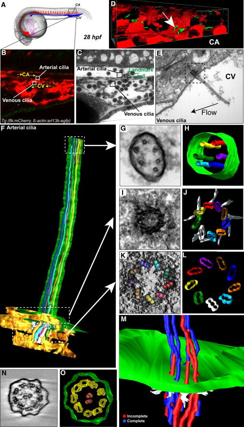
Fig. 2
Early Embryonic Vessels Are Ciliated
(A) Global view of a 28 hpf embryo.
(B) Single confocal section of the region of interest in the CA; endothelium is in red, primary cilia are in green. Boxes define two cilia of interest located in the CA (arterial) and CV (venous). Flow direction is indicated. See also Movie S8.
(C) Transmission electron microscopy (TEM) image of the region shown in (B).
(D) 3D reconstruction of the region of interest (arrow points to cilium of interest).
(E) TEM image of the venous cilia highlighted in (B) and (C).
(F–M) Electron tomography of the arterial cilia outlined in (B)–(D).
(F) Reconstruction of the portion of the protruding cilia near the cell surface.
(G–L) Single tomograms and modeling of regions located in the axoneme (G and H) and the basal body (I–L) are presented. Microtubules were color-labeled along the whole cilia. Note that the light blue microtubule is incomplete over the tomogram presented in (G) and (H). See also Movies S8, S9, and S10.
(M) A portion of the cilia proximal to the plasma membrane. Microtubules that disappear along the cilia axis are labeled in red. See also Movie S11.
(N and O) Single tomogram and modeling of a motile cilia located in the pronephros. Note the characteristic 9+2 microtubule doublet architecture.
See also Figure S2 and Movie S12.