
|
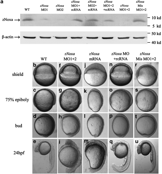
Fig. 2 Altered expression of zNoxa results in deformed embryos. (a) Protein was extracted from embryos at the shield stage and 30 µg of each sample was immunoblotted for zNoxa and β-actin. β-Actin served as an internal control. (b-u) The WT embryos (b-e), the zNoxa MO1+2-injected embryos (f-i), the zNoxa mRNA-injected embryos (j-m), the zNoxa MO1+2 plus rcRNA-injected embryos (n-q) and the zNoxa mismatch MO1+2-injected embryos (r-u) were shown at shield stage, 75% epiboly stage, bud formation stage and 24 h.p.f. WT and the injected mismatch-MO embryos were identical in appearance. Lateral views of embryos at shield and 75% epiboly stages, animal pole toward the top and dorsal to the right. Lateral views of embryos at bud and 24 h.p.f. stage, antetior toward the top and dorsal to the right