
|
Fig. S4
Histological features of dysplasia and HCC caused by UHRF1 over expression. (Related to Figure 4)
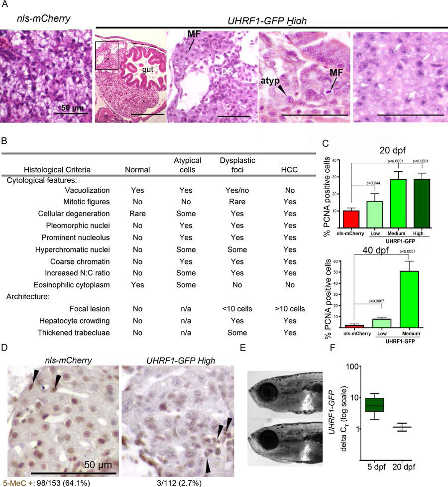
(A) H&E sections of UHRF1-GFP High livers in 20 dpf fish illustrate features of dysplasia and HCC that are not present in nls-mCherry controls. Features represented in the images in A include hepatocyte crowding, ductular reaction (DR), presence of mitotic figures (MF), atypical cells (atyp) and enlarged senescent cells (indicated by white arrows). The two areas marked by * in the far left panel of UHRF1-GFP High fish indicate two separate lesions in the sameliver. The boxed region is enlarged in the panel on the right. (B) Histological criteria including cytological and architectural changes used to define features of atypical cells, dysplastic foci and HCC in zebrafish. (C) PCNA positive and negative hepatocytes were counted in >200 hepatocytes from 2-8 fish per cohort of Tg(fabp10:nls-mCherry) and UHRF1-GFP overexpressing lines at 20 dpf and 40 dpf. Error bars are SD. (D) 20 dpf Cherry controls and UHRF1-GFP High sections were stained with anti-5MeC and the percent of 5MeC stained hepatocytes is shown. Arrows point to positively staining cells that are not hepatocytes. (E) Two UHRF1-GFP High fish were randomly selected on 20 dpf to image GFP in the liver as a measure of transgene expression. (F) qRT-PCR analysis of UHRF1 mRNA expression at 5 and 20 dpf normalized to rpp0 demonstrates that transgene expression is maintained, albeit decreased, in older UHRF1-GFP High fish. Boxes represent 75th and 25th percentile, bars indicate lowest and highest values.
Reprinted from Cancer Cell, 25(2), Mudbhary, R., Hoshida, Y., Chernyavskaya, Y., Jacob, V., Villanueva, A., Fiel, M.I., Chen, X., Kojima, K., Thung, S., Bronson, R.T., Lachenmayer, A., Revill, K., Alsinet, C., Sachidanandam, R., Desai, A., SenBanerjee, S., Ukomadu, C., Llovet, J.M., and Sadler, K.C., UHRF1 overexpression drives DNA hypomethylation and hepatocellular carcinoma, 196-209, Copyright (2014) with permission from Elsevier. Full text @ Cancer Cell