
|
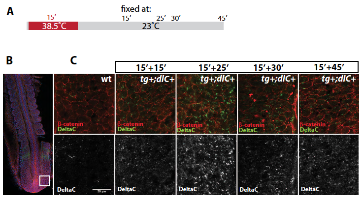
Fig. S1
Time course of DeltaC protein expression in tg+;dlC+ transgenic embryos after a single heat-shock. 15 hpf embryos were heat-shocked at 38.5 ºC for 15 minutes and left to recover at 23ÚC for varying lengths of time before fixation. (A) Scheme of the treatment. (B) Flat-mounted embryo, dorsal view; white box shows region of posterior PSM selected for analysis. (C) Enlargements of boxed region marked in B, for a series of embryos fixed at the indicated times and immunostained for DeltaC (green) and beta-catenin (red, marking plasma membrane); confocal imaging. Levels of cell-surface-associated DeltaC have begun to increase at 15+15 minutes, reach a peak at 15+25 minutes, and have declined markedly by 15+30 minutes. Pictures are representative of 5 transgenic embryos analysed by confocal microscopy at each time point.