
Image
|
Figure Caption
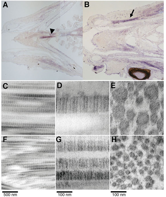
Fig. 3 Expression and ultrastructural analysis of adult zebrafish craniofacial tendons and ligaments. (A,B) Coronal sections of juvenile zebrafish show tnmd expression near the sternohyoideus attachment (arrowhead) and in lateral regions attached to the mandible (arrow). tnmd is also detected in the perichondrium. Transmission electron microscopy of ligament (C,D) and tendon (F,G) longitudinal sections reveal the parallel arrangement of collagen fibrils with characteristic periodicity. Ligament (E) and tendon (H) cross-sections reveal round collagen fibrils.
Figure Data
Acknowledgments
This image is the copyrighted work of the attributed author or publisher, and
ZFIN has permission only to display this image to its users.
Additional permissions should be obtained from the applicable author or publisher of the image.
Full text @ Development