
|
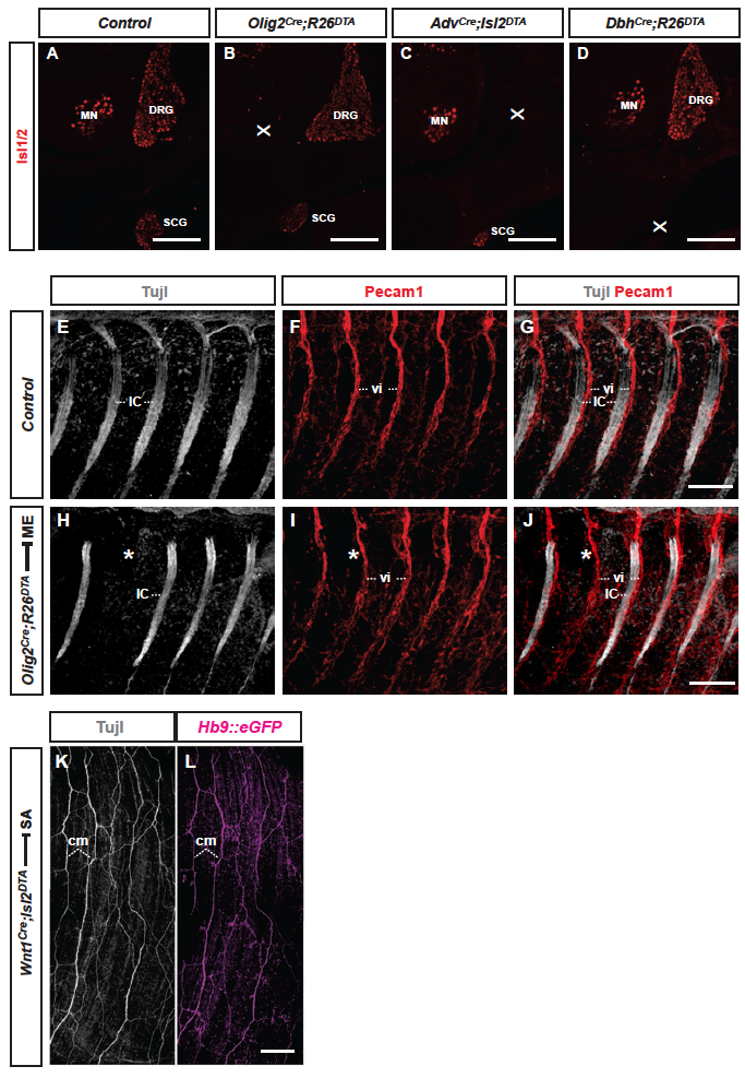
Fig. S6 Selective manipulation of peripherally projecting neuron types in mouse and relationship between PNs and vasculature. (A-D) Transversal section of E14.5 mouse embryos at trunk levels: anti-Isl1/2 immunofluorescence visualizes nuclei of motor neurons (MNs) in spinal cord, DRG and SCG neurons in control embryo (A), selective ablation of MNs in Olig2Cre;Rosa26lxstopDTA (B), selective ablation of DRG neurons in AdvCre;Isl2lxstopDTA (C), selective ablation of SCG neurons in DbhCre;Rosa26lxstopDTA (D). (E-G) Lateral wholemount view of E14.5 mouse trunk: spatial arrangement of intercostal PNs (IC, grey) (E,G) and intersegmental blood vessels (vi, red) (F,G). (H-J) Lateral wholemount view of E14.5 trunk in Olig2Cre;Rosa26lxstopDTA mouse embryo: intermittent absence of intercostal nerves (IC) (H,J) does not affect formation of intersegmental blood vessels (vi) (I,J). (K-L) Dorsal wholemeount view of AdvCre;Isl2lxstopDTA E18.5 mouse embryo: subdermal cutaneous maximus (cm) ME projections labeled by pan-axon marker anti-Tuj-1/βIII-tubulin (grey) and ME marker Hb9MN::eGFP. Scale bars: 150 µm in A-D; 300 µm in G,J,L.