
Image
|
Figure Caption
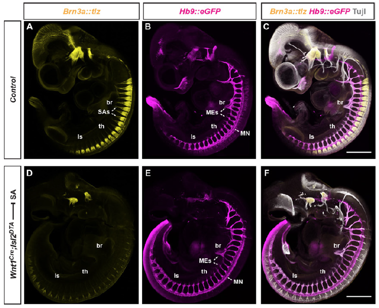
Fig. S5 Selective ablation of DRG neurons and SAs in mouse. (A-C) Lateral wholemount view of E10.5 mouse control embryo: normal appearance of peripheral SAs (yellow: Brn3atlz), MEs (magenta: Hb9MN::GFP) and pan-axon label (grey: anti-Tuj1 immunofluorescence). (D-F) Near-absence of DRG neurons (D,F), but not MNs or MEs (E,F) in Wnt1Cre;Isl2lxstopDTA embryo. Note: aberrant head structures, likely due to cranial neural crest defects. Abbreviations: br (brachial), th (thoracic), ls (lumbosacral).
Acknowledgments
This image is the copyrighted work of the attributed author or publisher, and
ZFIN has permission only to display this image to its users.
Additional permissions should be obtained from the applicable author or publisher of the image.
Full text @ Development