
|
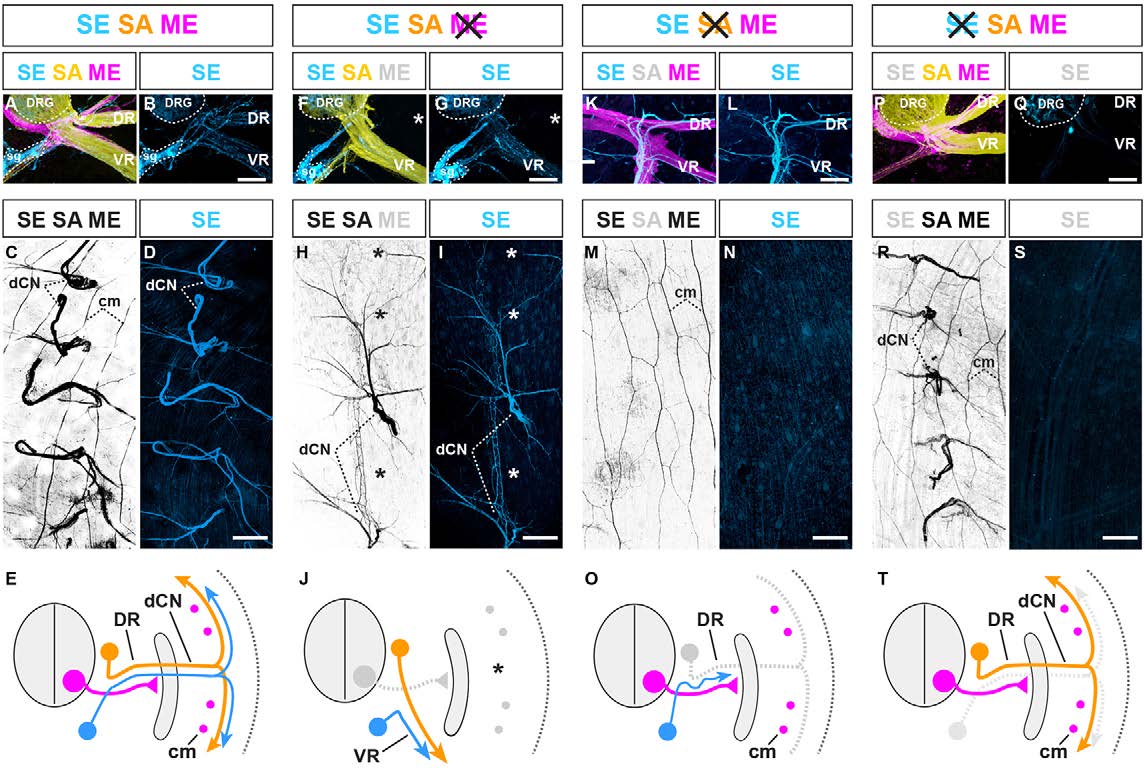
Fig. 7 SE trajectories are configured by pre-extending SAs and MEs. (A,B) Transverse section of E14.5 mouse embryo: SE (blue), SA (yellow) and ME (magenta) axons extending into dorsal (DR) and ventral (VR) nerve rami. (C) Whole-mount view of dorsal cutaneous nerve (dCN) axons fanning out into trunk dermis. (D) Visualization of SE axons only in same specimen. (E) Summary: three axon types in DR (VR, visceral or vascular trajectories are not depicted for simplicity). Magenta dots indicate cross-sectioned longitudinally projecting cutaneous maximus (cm) MEs. (F,G) Loss of dorsal (asterisk) and ventral misrouting of SA projections in the absence of MEs (OligCre;Rosa26fxstopDTA) (F) mirrored by SE (G) (asterisk). The converse dorsal misrouting of ventral SAs observed upon ME removal is not shown for simplicity. (H,I) Intermittent loss of dCNs (asterisks) and aberrant pattern of SA projections in the absence of MEs is mirrored by SEs (I). (J) Summary of F-I. (K-N) Initial peripheral extension of SEs along MEs in the absence of SAs (AdvCre;Isl2fxstopDTA) (note the higher degree of SE fasciculation, compared with control), but failure of SEs to innervate dermis (M,N) (remaining axons in M are subdermal cm MEs). (O) Summary of K-N. (P-Q) Normal appearance of ME, SA trajectories in the absence of SEs (DbhCre;Rosa26fxstopDTA). (R,S) Normal appearance of dorsal cutaneous nerves in the absence of SEs. (T) Summary of appearance of P-S. All images are representative of at least five embryos per condition. Scale bars: 100µm in B,G,L,Q; 300µm in D,I,N,S. sg, sympathetic ganglion.