
|
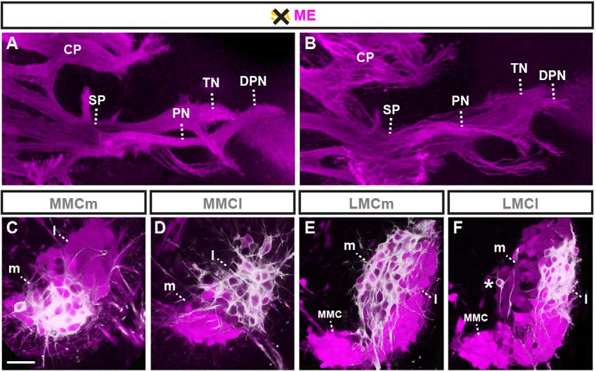
Fig. 4 SAs influence ME fasciculation but not trajectory or target choice. (A,B) Dorsal whole-mount view: MEs extending into hindlimbs in control (A) and in the absence of SAs in Wnt1Cre;Rosa26lxstopDTA mouse embryos (B). CP, crural plexus; DPN, deep peroneal nerve; PN, peroneal nerve; SP, sciatic plexus; TN, tibial nerve. (C-F) Transverse section of E12.5 thoracic (C,D) and lumbar (E,F) motor columns (magenta indicates Hb9MN::GFP): retrograde DiI tracing does not detect aberrant ME targeting at the level of columnar divisions. (C) Retrograde DiI tracing from epaxial muscle labels medial division of medial motor column (MMC). (D) Retrograde DiI tracing from hypaxial muscle labels lateral MMC. (E) Retrograde DiI tracing from ventral hindlimb labels Hb9::eGFPlow medial division of lateral motor column (LMC). (F) Retrograde DiI tracing from dorsal hindlimb labels Hb9MN::GFPhigh lateral LMC (asterisk indicates a possible Hb9MN::GFPhigh LMCl neuron in the process of lateral migration). Scale bars: 300µm in A,B; 50µm in C-F.