
|
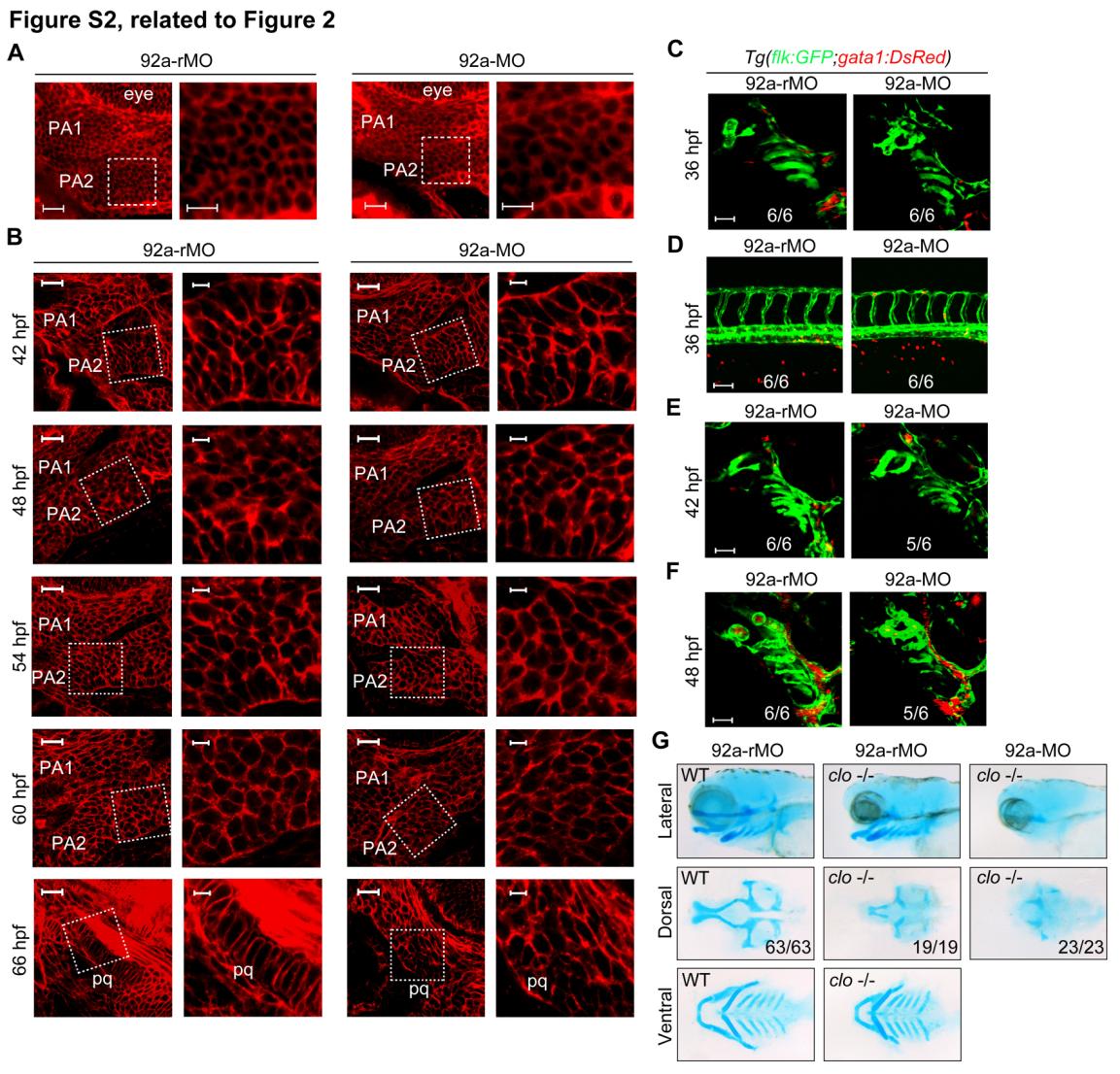
Fig. S2 mir92a inactivation impairs pharyngeal cartilage formation independent of blood flow, related to Figure 2. (A) Detection of cell surface proteins in the pharyngeal mesenchymal condensations by Peanut Agglutinin (PNA) staining. Embryos were injected with 4 ng 92a-rMO and 92a-MO, and collected for PNA staining at 60 hpf. The anterior first two pharyngeal arches (PA) were shown. The boxed area in the left image was presented at higher magnification in the corresponding right image. Scale bars, 20 µm (left) or 10 µm (right). (B) F-actin-labeled morphological changes in mir92a morphants at different stages. Wild-type embryos were injected with 4 ng 92a-rMO or 92a-MO at the one-cell stage and fixed at indicated stages for Phalloidin staining. Stained embryos were imaged by confocal microscopy with focus on the pharyngeal arch region. The order of the pharyngeal arches (PA) in the focus plane was indicated. For each panel, the boxed area in the left image was presented at higher magnification in the corresponding right image. At least 5 embryos were analyzed each group. Scale bars, 20 µm for the left image of each panel and 5 µm for the right image of each panel. (C-F) Confocal images of blood vessels (green) and blood cells (red) in the branchial (C, E, F) and trunk (D) regions. Tg(flk1:EGFP);Tg(gata1:DsRed) double transgenic embryos were injected with 4 ng 92a-rMO or 92a-MO at the one-cell stage and observed by confocal microscopy at 36 hpf, 42 hpf and 48 hpf. Scale bars, 50 µm. (G) Head skeleton of cloche mutant embryos. The embryos collected from clo m378 heterozygote intercrosses were injected with 4 ng 92a-rMO or 92a-MO at the one-cell stage. clo mutants were sorted out at about 48 hpf based on the absence of blood cells and stained with Alcian blue at 80 hpf. The images were orientated with anterior to the left.
Reprinted from Developmental Cell, 24(3), Ning, G., Liu, X., Dai, M., Meng, A., and Wang, Q., MicroRNA-92a upholds Bmp signaling by targeting noggin3 during pharyngeal cartilage formation, 283-295, Copyright (2013) with permission from Elsevier. Full text @ Dev. Cell