
|
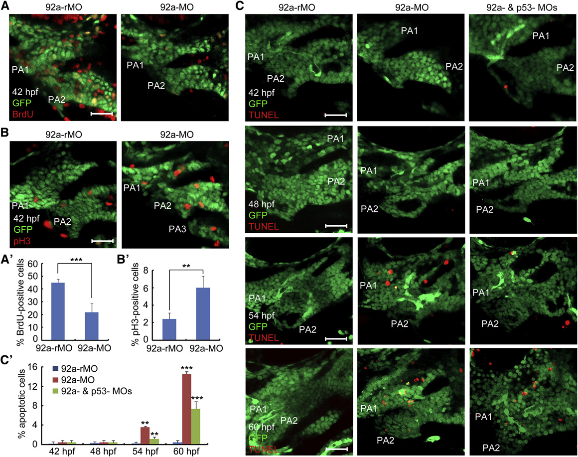
Fig. 3 Inhibition of mir92a Activity Impairs Proliferation and Survival of Chondrogenic Progenitors
(A–B2) Representative confocal sections showing BrdU- or pH3-positive cells (red) in the pharyngeal region. 92a-rMO or 92a-MO-injected (4 ng) Tg(fli1:EGFP) transgenic embryos were coimmunostained with anti-GFP and anti-BrdU (A) or anti-pH3 antibodies at 42 hpf (B). The images shown were lateral views for the first two pharyngeal arches (PA1 and PA2) (similarly labeled in C). Scale bars, 20 µm. The percentage of BrdU- or pH3-positive cells among GFP-positive pharyngeal chondrogenic progenitors, based on five embryos (one to two images each) for each group, were shown in (A2) and (B2). p < 0.01; p < 0.001.
(C and C2) Detection of apoptotic cells in the pharyngeal regions. Tg(fli1:EGFP) transgenic embryos were injected with 92a-rMO, 92a-MO, or 92a-MO plus p53-MO (at 4 ng each) and collected at indicated stages for TUNEL assay. Representative images were shown in (C). The ratio of TUNEL- and GFP-positive cells (orange) to GFP-positive cells in the first and second pharyngeal arches were calculated from five embryos with one to two images each and presented in (C). Scale bars, 20µm.
See also Fig. S3.
Reprinted from Developmental Cell, 24(3), Ning, G., Liu, X., Dai, M., Meng, A., and Wang, Q., MicroRNA-92a upholds Bmp signaling by targeting noggin3 during pharyngeal cartilage formation, 283-295, Copyright (2013) with permission from Elsevier. Full text @ Dev. Cell