
|
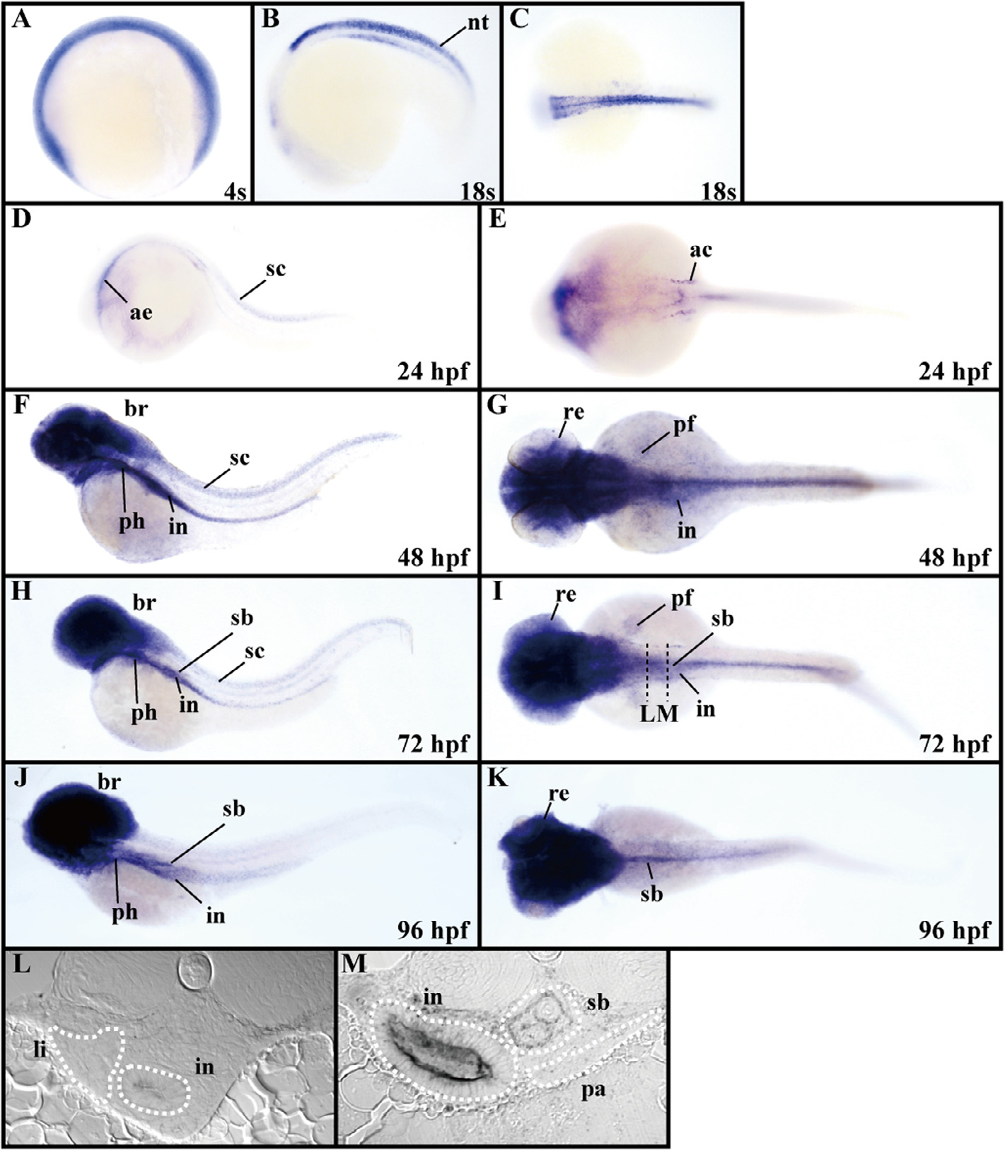
Fig. 2 Expression patterns of dnmt3ab in zebrafish embryos and larvae. (A–K) dnmt3ab expression was examined by whole-mount in situ hybridization at the 4s and 18s stages, and at 24, 48, 72, and 96 hpf. Lateral views, anterior to the left (A, B, D, F, H, and J). Dorsal views, anterior to the left (C, E, G, I, and K). (L and M) Transverse sections showing dnmt3ab expression. The transverse sections were cut at the levels indicated by the dashed black lines in (I). ac, auditory capsule; ae, anterior endoderm; br, brain; in, intestine; nt, neural tube; li, liver; pa, pancreas; pf, pectoral fin buds; ph, pharyngeal arches; re, retina; sb, swim bladder; sc, spinal cord.
Reprinted from Gene expression patterns : GEP, 14(2), Takayama, K., Shimoda, N., Takanaga, S., Hozumi, S., and Kikuchi, Y., Expression patterns of dnmt3aa, dnmt3ab, and dnmt4 during development and fin regeneration in zebrafish, 105-110, Copyright (2014) with permission from Elsevier. Full text @ Gene Expr. Patterns