
Fig. 6
- ID
- ZDB-IMAGE-140515-41
- Publication
- Sun et al., 2014 - Extraembryonic signals under the control of MGA, Max, and Smad4 are required for dorsoventral patterning
- All Figures
- Figures for Sun et al., 2014
|
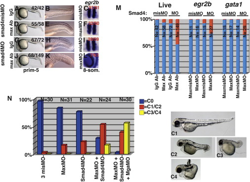
Fig. 6
MGA, Max, and Smad4 Are Required Synergistically in the YSL
Embryos were injected into the YSL with suboptimal doses of smad4misMO (A–F, M, and N) or smad4MO (G–N) along with 100 pg of IgG (A, B, G, H, and M), 100 pg anti-Max antibody (D, E, J, K, and M), maxmisMO (C, I, M, and N), maxMO (F, L, M, and N), and mgamisMO or mgaMO (N). One nanogram each of MO was injected, except for 2 ng each of MO in (C), (F), (I), (L), and (N).
(A, B, D, E, G, H, J, and K) Images of live morphants.
(C, F, I, and L) egr2b expression at 8-somites.
(M) Frequency of tailfin defects, expanded egr2b expression or expanded gata1 expression (red).
(N) Embryos injected with 6 ng MO total (2 ng each MO) and scored at Long-pec according to the dorsoanterior index (Mullins et al., 1996). The key is to the right, and representative embryos from each class are depicted. Due to limited space, only experimental MOs are listed on the x axis, but in each case the applicable control MOs were coinjected so that the total MO dose remained constant.
Reprinted from Developmental Cell, 28(3), Sun, Y., Tseng, W.C., Fan, X., Ball, R., and Dougan, S.T., Extraembryonic signals under the control of MGA, Max, and Smad4 are required for dorsoventral patterning, 322-334, Copyright (2014) with permission from Elsevier. Full text @ Dev. Cell