
|
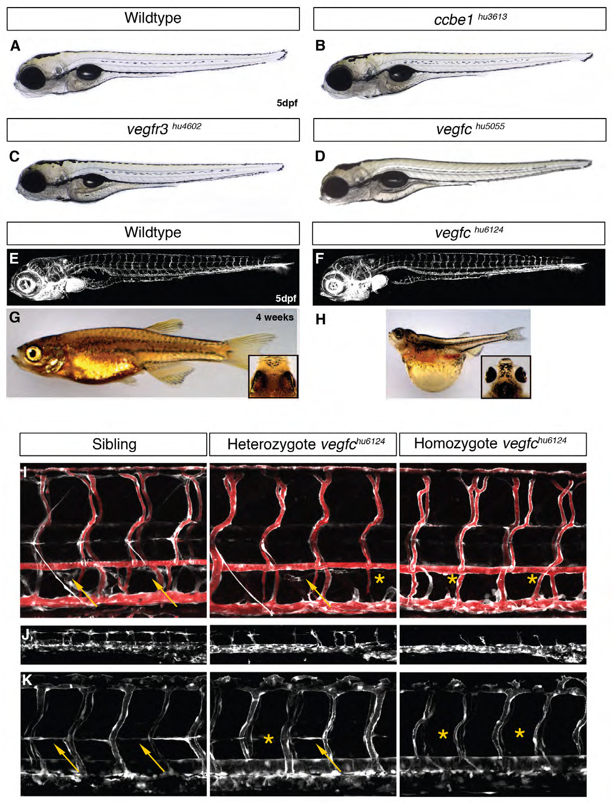
Fig. S1
Morphology of vegfc, ccbe1 and vegfr3 mutants and additional phenotypes of vegfc mutants. (A-D) Mutant hu5055 that specifically lacks lymphatic vasculature (Figure 2F and 2H) (D) is undistinguishable from vegfr3 (C) and ccbe1 (B) mutants. (E-H) Gross morphology of vegfc mutants. Confocal projections in the Tg(fli1a:EGFP) background. At 5 dpf, vegfchu6124 homozygote mutant display a specific loss of the lymphatic vasculature with apparently normal blood vasculature (F), as compared to wildtype (E). Mutant embryos that survive after 5dpf, display severe edema in the trunk area, as well as around the eyes (H) compared with wildtype siblings (G). (I-K) Phenotypes of heterozygous and homozygous vegfc mutants. (I) Angiographies in Tg(fli1a:EGFP) transgenic wildtype sibling, vegfchu6410 heterozygote and homozygote mutant embryos reveal the presence of blood flow and thoracic duct (TD) defects in mutants. In the vegfchu6410 early stop allele, heterozygote mutant embryos display a partial loss of TD development while in homozygotes no TD is observed. (J) Secondary sprouting in plcg1 morphants. In plcg morphants, arteries are missing facilitating the visualization of venous sprouts. As compared to wildtype, only a few secondary sprouts are observed in heterozygote mutants, and even fewer in homozygote mutant embryos. (K) PLs in a wildtype sibling, vegfchu6410 heterozygote and homozygote mutant embryos. As compared to wildtype, heterozygote embryos develop less PLs. In homozygote vegfchu6410 mutants no PLs are present.