
|
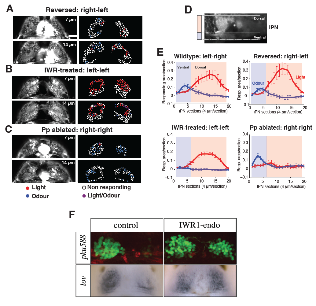
Fig. S2
Functional responses to visual and odour stimulations in fish with reversed, double-left and double-right epithalami in the Hb and IPN.
(A-F) Raw images of two z-planes (at 7 and 14 μm depth) of the dorsal Hb of 4 dpf Tg(elavl3:GCaMP5G) reversed (A), double-left (B), and double-right (C) brained fish (left panels). In the right panels, dHb neuron cell bodies, indicated as regions of interests (ROIs), are colour-coded in red, blue, violet, or white dependent upon their response to light, odour, both, or neither respectively. In the case of parapineal reversal Tg(elavl3:GCaMP5G) fish were crossed with the Tg(foxD3:GFP) fish to visualize the parapineal on the right side (A). For the Parapineal (Pp) ablation, Tg(elavl3:GCaMP5G) fish were crossed with Tg(foxD3:GFP) and Tg(flh:GFP) fish (C). Scale bar 20 μm. (D) Sagittal view of the IPN highlighting the dorsal (light red) and ventral (light blue) IPN. (E) Depth profile of averaged light (red line) and odour (blue) IPN responses from dorsal to ventral in wildtype, reversed, double-left, and double-right conditions. Red and blue lines are averages of three trials for each stimulus modality. (n=7 wildtype, n=6 reversed, n=8 double-left, and n=6 doubleright). (F) Dorsal views of the epithalami of control (left) and IWR1 treated (right) embryos showing expression of markers normally lateralized to the majority of left habenular neurons. Top shows the pku588 transgene (Lu Wen, Bo Zhang, Shou Lin, MC and SW, unpublished data) and B,B′ shows kcdt12.1 (Gamse et al., 2003) expression. IWR1-mediated suppression of Wnt-signalling leads to many right-sided habenular neurons expressing markers typical of the neurons on the left. This consequently leads to an overtly double-left phenotype.