
Image
|
Figure Caption
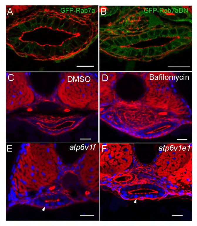
Fig. S3
The degradation pathway is not involved in lumen formation. (A,B) Confocal cross sections from Tg(hsp70l:GFP-Rab7) and Tg(hsp70l:GFP-Rab7DN) embryos. Phalloidin (red). (C,D) Confocal cross sections from DMSO and bafilomycin treated embryos. (E,F) Confocal cross sections from atp6 v1f and atp3v1e mutant embryos. Phalloidin (red). Scale bars: 20 μm.
Acknowledgments
This image is the copyrighted work of the attributed author or publisher, and
ZFIN has permission only to display this image to its users.
Additional permissions should be obtained from the applicable author or publisher of the image.
Full text @ Development