
Fig. SX.9
- ID
- ZDB-IMAGE-140509-7
- Publication
- Venkatesh et al., 2014 - Elephant shark genome provides unique insights into gnathostome evolution
- All Figures
- Figures for Venkatesh et al., 2014
|
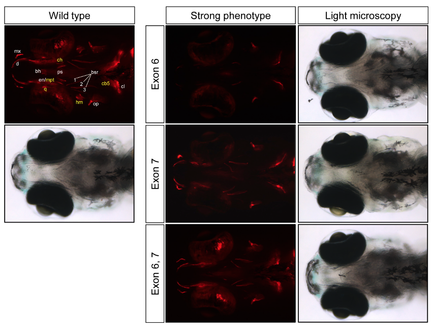
Fig. SX.9
Targeted mutagenesis of zebrafish spp1 by sgRNA:Cas9 results in reduced bone formation in 15 dpf embryos.
Left panel: ventral view of a 15 dpf wild type embryo stained with Alizarin red to reveal sites of bone deposition (red fluorescence). Its light microscopy image is given below. Endochondral bones (cb5, ceratobranchial 5; ch, ceratohyal; hm, hyomandibula; mpt, metapterygoid; q, quadrate) are highlighted in yellow whereas dermal bones (bh, basihyal; bsr, branchiostegal ray; cl, cleithrum; d, dentary; en, entopterygoid; mx, maxilla; op, operculum; ps, parasphenoid) are highlighted in white. Entopterygoid (en) and metapterygoid (mpt) are fused and hence cannot be readily distinguished. However, both are affected in spp1 mutants shown in the right panel. Right panel: ventral views of 15 dpf embryos injected with Cas9 mRNA together with single guide RNA (sgRNA) targeting spp1 exon 6, exon 7 or both exons. The ‘strong’ bone-reduction phenotype embryos are shown (stained with Alizarin red). Light microscopy images of the mutants show normal development of the embryos.