
Fig. SX.4
- ID
- ZDB-IMAGE-140509-3
- Publication
- Venkatesh et al., 2014 - Elephant shark genome provides unique insights into gnathostome evolution
- All Figures
- Figures for Venkatesh et al., 2014
|
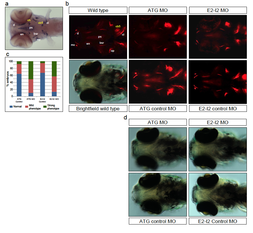
Fig. SX.4
Reduction of bone deposition in spp1 zebrafish morphants.
(a) spp1 is specifically expressed in cells surrounding the bone matrix. Ventral view of a 5 dpf embryo hybridized with spp1 RNA probe. Endochondral bones (ch, ceratohyal; cb5, ceratobranchial 5) are highlighted in yellow whereas dermal bones (bsr, branchiostegal ray; cl, cleithrum; d, dentary; en, entopterygoid; op, operculum; ps, parasphenoid) are highlighted in white. (b) Ventral views of 5 dpf wild type, ATG MO, ATG control MO, E2-I2 MO and E2-I2 control MO embryos. Embryos were stained with alizarin red to reveal sites of bone deposition and imaged under red fluorescence. For one wild type embryo (bottom left), a merged bright field image and red fluorescence signal is shown to simultaneously visualize anatomical structures and bone deposition. (c) Proportion of embryos showing normal phenotype (resembling wild type), ‘mild’ bone-phenotype and ‘strong’ bone-phenotype (the latter showing the most reduction of bones). ATG MO and E2-I2 MO morphants show a significantly higher proportion of strong bone-phenotype (p<0.01, Fisher’s exact test)compared to their respective controls. (d) Bright field images of embryos shown in panel (b) showing normal growth of morphant embryos.