
|
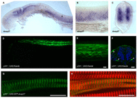
Fig. 4
dusp27 is expressed during embryonic muscle development. (A–C) In situ hybridization for dusp27 expression at 24 hpf. Prominent expression is seen in the somites (asterisk). (B) Lateral view above the yolk extension. (C) Cross-section through the trunk caudal to the yolk extension. (D–F) Kaede reporter expression in y241; UAS:kaede embryos. (D) Kaede expression is strongly expressed in trunk muscle at 24 hpf and (E) continues to label myofibers in 6 dpf larvae. Scale bar: 50 μm. (F) Early expression in somites is observed at 15 somites in a small number of cells adjacent to the notochord (arrowheads), as well as at the lateral edge of the somite (asterisk). Scale bar: 50 μm. (G,H) Expression of GFP-Dusp27 in muscle cells shows a striated pattern (green, G) that alternates with α-actinin (red in merge, H). Scale bars: 10 μm.