
|
Fig. S3
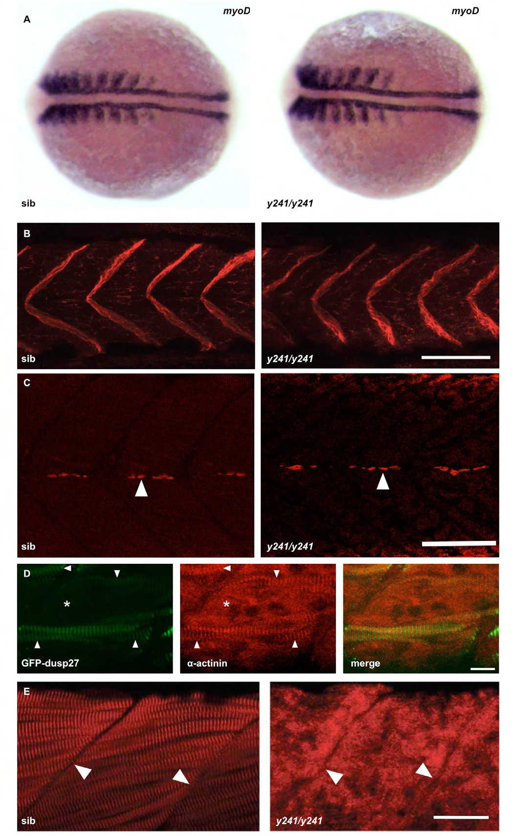
Additional analysis of muscle markers in y241 mutants. (A) In situ hybridization for myoD expression in 10 somite sibling (left) and y241 mutant (right) embryos. After photographing, embryos were genotyped by PCR. (B) Immunofluorescence staining for dystrophin (merged z-stack through muscle) shows similar localization at somite boundaries in sibling and y241 mutants in 48 hpf embryos. Scale bar 50 μm. (C) 4D9 staining of engrailed expression in muscle pioneer cells indicates the presence of grossly normal densities of pioneer cells (arrows). Scale bar: 50 μm. (D) In y241 mutants injected with the GFP-dusp27 plasmid, muscle fibers expressing GFP (left panel, arrowheads) recover a normal striated pattern of α-actinin expression (middle panel) while regions of the somite without GFP expression do not show patterned α-actinin expression (asterisk). Merged view on right. Scale bar 10 μm. (E) The normal striated pattern of ryanodine receptor staining is disrupted in 48 hpf y241 mutant embryos. Arrowheads point to somite boundaries. Scale bar 25 μm.