
|
Fig. S2
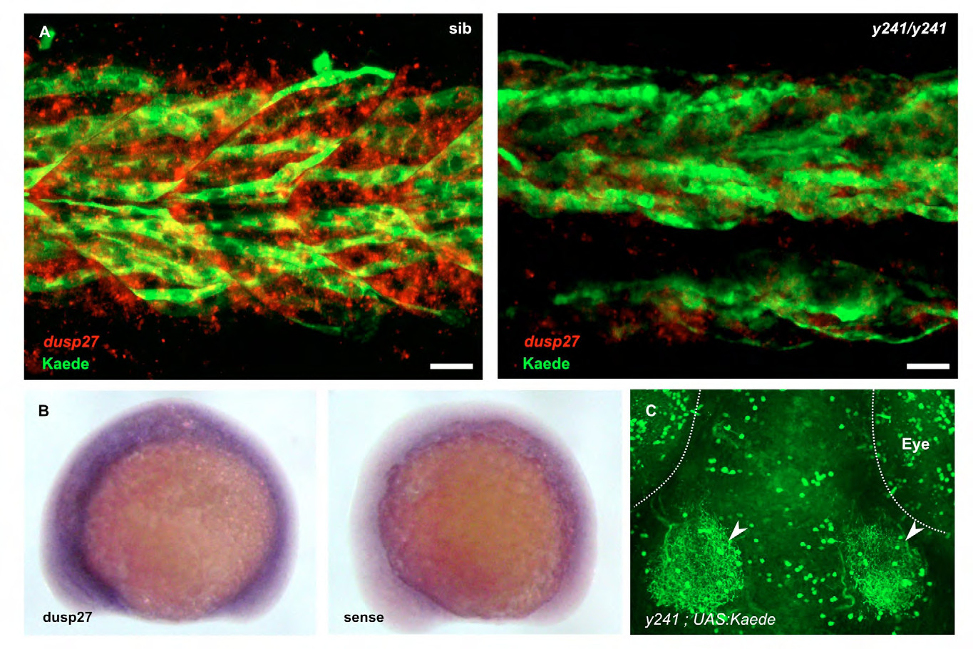
Additional analysis of dusp27 expression. (A) Muscle fibers labeled by anti-kaede staining (green) in y241 ; UAS:Kaede heterozygotes (left) at 30 hpf co-express dusp27 (red, in situ hybridization), while dusp27 expression in mutants is strongly diminished. Note that the expression of dusp27 in fibers not labeled by kaede is likely due to variegated expression of the UAS:Kaede transgene, a commonly observed phenomenon in zebrafish UAS lines (Goll et al., 2009). Scale bar 20 μm. (B) in situ hybridization for dusp27 at 15 somites (right panel shows sense control) (C) In 6 dpf brain, Kaede prominently labels deep tectal neurons that extend neurites into the tectal neuropil (arrowheads) as well as neuronal groups in the retina, cerebellum and hypothalamus (not shown). Scale bar 50 μm.