
|
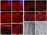
Fig. 7
Fast muscle myofibrils are disrupted in y241 mutants. (A–E) Immunofluorescence in fast-twitch fibers in 48 hpf sibling and y241 mutant embryos. (A) F310 labeling of fast-twitch-fiber myosin shows disruption of thick filaments. Scale bar: 25 μm. Insets show rotated transverse view through the somite (merged view of DAPI in blue and F310 in red). (B) CH1 labeling of tropomyosin shows disruption of thin filaments. Scale bar: 20 μm. (C) α-actinin staining shows loss of Z-lines in fast-twitch fibers. Scale bar: 25 μm. (D) Anti-titin staining. Scale bar: 30 μm. (E) DHPR in fast-twitch fibers. Scale bar: 25 μm. (F) Transmission electron microscopy in myofibers of 3 dpf y241 siblings and mutants, showing Z-lines (arrows) and triads (arrowheads). Scale bars: 500 nm.