
Image
|
Figure Caption
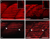
Fig. 6
Slow muscle myofibrils are partially disrupted in y241 mutants. (A) F59 staining against slow-twitch muscle myosin heavy chain in 48 hpf embryos shows the presence of slow muscle fibers at the lateral edge of the somite in both mutants and siblings but disorganized thick filaments in y241 mutants. Scale bar: 20 μm. Insets show rotated transverse view through the somite. (B) α-actinin staining in slow-twitch fibers shows preserved Z-lines (arrowheads) in y241 mutants. Scale bar: 25 μm.
Figure Data
Acknowledgments
This image is the copyrighted work of the attributed author or publisher, and
ZFIN has permission only to display this image to its users.
Additional permissions should be obtained from the applicable author or publisher of the image.
Full text @ Dis. Model. Mech.